Contrast-enhanced (CE) mammography might be better suited than contrast-enhanced MRI for confirming some cases of suspicious breast cancer -- especially when radiation dose-reducing protocols are used, according to a study published February 14 in the Journal of Magnetic Resonance Imaging.
In a direct comparison of the modalities, low-dose CE mammography significantly outperformed CE-MRI in specificity and positive predictive value (PPV) and matched the latter modality with a high detection rate for malignant lesions. Given the results, low-dose CE mammography might reduce the number of unnecessary biopsies.
"In addition, due to its high lesion detection and the challenge of characterizing small and nonmass enhancements, CE-MRI often leads to biopsies for lesions that prove to be benign," wrote the authors, led by Dr. Paola Clauser from the Medical University of Vienna in Austria (J Magn Reason Imaging, February 14, 2020). "Low-dose CE mammography could be a cost-effective alternative and might help to improve the management of patients with suspicious or inconclusive findings on mammography, tomosynthesis, or ultrasound."
To radiate or not?
Contrast-enhanced MRI has often been used as the first follow-up option when mammography and/or ultrasound cannot definitely detect or diagnose cases of suspected breast cancer and to screen women for the disease. While the modality offers high sensitivity, it also costs more, has a longer scan time, and often requires additional workup, compared with other imaging options.
But recent studies evaluating CE mammography show that it can be quite sensitive for breast cancer detection as well and is "very well accepted by patients," Clauser and colleagues noted. The downside of CE mammography is greater radiation exposure to patients, compared with full-field digital mammography and potentially digital breast tomosynthesis. That radiation load, however, could be reduced by removing the antiscatter grid and adding a software-based scatter-correction method to ensure good image quality.
In the balance of risk versus benefit, is it worth exposing patients to radiation with CE mammography to increase the chances of detecting breast cancer? Can comparable results be achieved with less radiation? Or is it best to stay with CE-MRI, which already has proved its efficacy with no radiation exposure?
"Up to now, only a few studies have compared CE mammography to CE-MRI, and most of those studies were retrospective," the authors wrote. "Nevertheless, the studies indicated that CE mammography has the potential to become an alternative to CE-MRI."
To help settle the debate, Clausen and colleagues initiated a prospective, single-center study that included 80 women (mean age, 54.3 ± 11.2 years) with suspicious results on conventional mammography, tomosynthesis, or ultrasound. All the participants underwent both imaging exams within 24 to 72 hours, with image-guided biopsies performed on the most suspicious lesion after both CE mammography and CE-MRI were completed.
Imaging protocols
CE mammography was performed on a modified full-field digital mammography (FFDM) device (Mammomat Inspiration, Siemens Healthineers). The protocol included both low- (28-32 kVp) and high-energy (49 kVp) images, which were acquired consecutively during a single breast compression.
To reduce radiation dose, the researchers omitted the antiscatter grid and added a dedicated, software-based scatter-correction method. An antiscatter grid was utilized for women with breasts with a thickness greater than 70 mm. In addition, the patients received a single dose of a nonionic iodine contrast agent (iobitridol/Xenetix 350, Guerbet), based on 2 mL/kg of body weight. The researchers obtained a total of 640 mammographic views.
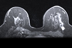
MRI scans were performed on either a 1.5- or 3-tesla scanner, with a protocol that included a T2-weighted sequence and a gradient-echo, T1-weighted sequence before and after a single dose of a gadolinium-based contrast agent.
Three experienced readers were asked to determine the presence of lesions, along with location, type, and size, and BI-RADS score. A BI-RADS score of 1 to 3 indicated a negative or nonsuspicious ruling, while BI-RADS 4 and 5 scores were considered positive or suspicious. Histology served as the gold standard.
"Only one lesion per breast was considered," the authors explained. "When more lesions were described, only the most suspicious lesion per breast, for which a histological verification was available, was considered."
In their review, the trio found 93 histologically verified breast lesions, of which 61 lesions (66%) were malignant and 32 lesions (34%) were benign. There were 67 breasts with no lesions. Lesion size ranged from 4 mm to 120 mm on lose-dose CE mammography and 4 mm to 100 mm on CE-MRI.
In comparing the results of the two modalities, the researchers found the overall lesion detection rate was significantly greater with CE-MRI than with low-dose CE mammography. Conversely, low-dose CE mammography significantly outperformed CE-MRI on specificity and positive predictive value (PPV). There were no statistically significant differences between the two modalities for sensitivity and negative predictive value (NPV), while low-dose CE mammography was comparable to CE-MRI for accuracy.
| CE-MRI vs. CE mammography for suspicious breast cancer | |||
| CE-MRI (%) | CE mammography (%) | p-value | |
| Detection rate | 92.5-94.6 | 79.6-91.4 | 0.014* |
| Specificity | 37.5-53.1 | 46.9-96.9 | 0.001* |
| PPV | 73.3-77.3 | 76.4-97.6 | 0.007* |
| Sensitivity | 83.6-93.4 | 65.6-90.2 | 0.086 |
| NPV | 63.0-76.5 | 59.6-71.4 | 0.780 |
| Accuracy | 72.0-75.3 | 75.3-76.3 | 0.514 |
So what do all the numbers mean? Given that low-dose CE mammography outperformed CE-MRI on specificity and PPV and showed a "high detection rate for malignant lesions," the modality "might help to reduce false-positive biopsies while increasing the cancer detection rate," Clausen and colleagues wrote. "This is of great advantage, as CE-MRI of the breast is a rather expensive examination."
The researchers also recommended larger studies to confirm if CE mammography is indeed a "safe alternative to breast CE-MRI in all patients."
Study disclosures
The study was supported by a grant from Siemens Healthineers and Guerbet. Clauser and colleagues stated they had full control of all data and statistical results.


