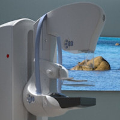
A women's imaging center in Florida has reported that spa-like rooms with soothing videos, scents, and sounds resulted in better mammogram image quality. Their study is the first to link multisensory environments to better breast images, according to the researchers.
Multisensory environments are thought to reduce patients' pain and anxiety by either overloading the brain's pain sensors or by providing a distraction. The authors believed this improvement in patient outcomes then translated to better mammogram image quality in their 300-person study, which was published on July 17 in the Journal of Breast Imaging.
"Essentially, the patients may experience less pain and anxiety. This may lead to improved positioning which influences image quality and provides a superior image to the radiologist for interpretation," wrote the authors, led by Shakira Sarquis-Kolber, director of women's imaging at the Boca Raton Regional Hospital's Lynn Women's Health & Wellness Institute.
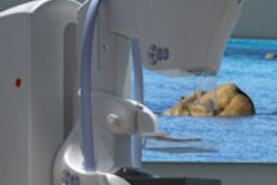
The retrospective analysis focused on a four-room facility in Boca Raton that performs 20,000 breast screenings per year. In 2014, the facility upgraded one of its mammography rooms to incorporate a SensorySuite room environment from GE Healthcare.
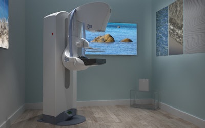 GE's SensorySuite concept upgrades mammography rooms with stimuli such as landscapes, aromatherapy, and soothing sounds.
The upgraded room featured a spa-like atmosphere, including soothing sounds and air infused with a light, citrus scent. Patients also selected an environmental scene to play on three wall displays.
The authors compared imaging data from 303 women who underwent annual mammography at the facility. In the first year, all patients visited a standard, plain-walled room, but in the second year, they underwent mammography in the upgraded room.
Although the rooms had different environments, they contained the same GE mammography system. Four mammography-certified techs performed all patient mammograms using the same procedures and protocols.
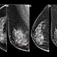
To compare the image quality from one year to the next, the authors used posterior nipple line (PNL) measurements, a commonly used indicator of image quality. A radiologist with a breast imaging specialty reviewed the measurements for accuracy.
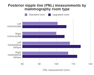
The PNL measurements were significantly greater for mammograms taken in the upgraded room than the standard one, with an average improvement of 5 mm to 6.2 mm in the multisensory environment. Notably, the image quality improved even without a significant difference in the compression force.
"This is the first study to identify improved image quality from the use of a mammography room with a multisensory environmental upgrade," the authors wrote.
The authors attributed the image quality improvement to a reduction in patients' fear and anxiety, which in turn improved positioning. Although the results are promising, future, multisite studies are needed to verify the findings.
"Future research using a multicenter study with larger sample sizes would allow for greater generalizability," the authors concluded.


