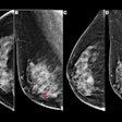
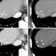
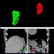
| FIGURE 2.1.6 Dysplasia and carcinoma in long-standing ulcerative colitis. Contrast-enhanced CT images (A to C) show a cecal carcinoma (B, asterisk) superimposed on features of long-standing UC, including a foreshortened, ahaustral appearance with prominence of the pericolonic vessels. BE image (D) from a second patient with long-standing UC shows irregular long-segment narrowing of the ascending colon from adenocarcinoma. Note again the foreshortened, ahaustral colon and findings suggestive of "backwash ileitis." Colonoscopy image (E) from a third UC patient shows a flat reddish lesion that demonstrated low-grade dysplasia on biopsy. Unfortunately, the patient was lost to follow-up and returned 2 years later with a malignant stricture in this region (F). |
Atlas of Gastrointestinal Imaging FIGURE 2.1.6 Dysplasia and carcinoma in long-standing ulcerative colitis
Latest in Home


