Minnies finalists, page 3
Scientific Paper of the Year
Patient-centered radiology: Where are we, where do we want to be, and how do we get there? Kemp JL et al, Radiology, June 20, 2017. To learn more about this paper, click here.

This research cuts to the heart of an existential dilemma that radiology is facing: how to improve the specialty's value in the healthcare continuum by becoming more visible to patients.
In the study, a research team led by Dr. Jennifer Kemp of Diversified Radiology in Colorado surveyed nearly 700 radiologists to gauge their attitudes toward spending more time with patients. Those surveyed generally agreed that radiologists should spend more time communicating with patients, with academic radiologists and those in subspecialties with more patient interaction finding more value in it.
So if radiologists agree that they should be interacting with patients, why aren't they actually doing it? Kemp and colleagues found a variety of reasons, ranging from time and workload demands to resistance from referring physicians who might view radiologists as encroaching on their turf.
The study exposes a "fundamental tension" between what radiologists are being told to do and what they actually have time to do, the researchers concluded. Because they are being paid to read images rather than talk to patients, radiologists may need to adapt to decreased income if they really want to practice more patient-centered care.
Women's awareness and perceived importance of the harms and benefits of mammography screening: Results from a 2016 national survey. Yu J et al, JAMA Internal Medicine, June 26, 2017. To learn more about this paper, click here.

One of the major criticisms that mammography skeptics raise against breast screening is that it exposes women to overdiagnosis, or the detection of cancers that might never pose a health risk. If women only knew about overdiagnosis, they would be less enthusiastic about screening, the theory goes.
This paper undercuts that argument. More than 400 women surveyed by researchers from the University of Minnesota rated the lifesaving benefits of mammography as being far more important to them than the anxiety and stress that might be caused by alleged "harms" produced by screening. And women who had been screened most recently had the most positive view of mammography.
Unfortunately, this wasn't quite the result the researchers were looking for. They speculated that women view mammography so positively due to a "lack of balance" from physicians, public health officials, and the news media, and that "targeted education and communication" is needed to change their opinions.
Best New Radiology Device
Somatom go line of CT scanners, Siemens Healthineers
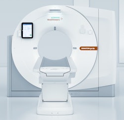 Somatom go.Up from Siemens.
The Somatom go line of CT scanners from Siemens Healthineers represents an intriguing shift in the development of CT instrumentation, with a focus on improving workflow in the CT suite rather than just boosting raw horsepower.
Available in 32-slice (go.Now) and 64-slice (go.Up) models, the Somatom go scanners are based on a tablet computer that enables technologists to operate the system with controls that duplicate those found on the scanner console.
This creates a mobile workflow that frees technologists from being tethered to the scanner, enabling them to spend more time closer to patients, according to the company.
Siemens believes that the mobile workflow made possible by the system will give customers more flexibility in siting the units, and it will enable them to enjoy lower operational costs -- for example, a dedicated operator's room is no longer necessary. Both systems use Siemens' Stellar CT detector technology, as well as the company's iterative reconstruction dose reduction protocols.
Introduced at RSNA 2016, the Somatom go systems received U.S. Food and Drug Administration (FDA) clearance in March 2017.
BodyTom Elite portable CT scanner, Samsung NeuroLogica
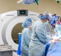 BodyTom Elite from Samsung.
Samsung NeuroLogica has made its name developing portable CT scanners that can be sited in cramped spaces such as operating rooms, moved about a hospital, or even installed in ambulances.
Introduced in July 2017 with 510(k) clearance, BodyTom Elite represents the latest iteration of the company's 32-slice BodyTom platform for full-body scanning. The upgraded unit includes a new visual design and upgraded software and hardware, according to the company.
The system includes a number of features designed to counteract issues that can occur when the scanner is moved, such as a tilt sensor to detect shifts in the floor and a step correction feature that makes adjustments in response to slight movements. A battery backup unit means the system can be transported without shutting down.
On the clinical side, BodyTom Elite includes a range of features found on more powerful scanners, such as low-dose lung cancer screening, metal artifact reduction, automatic exposure control, and 30 new custom kernels to give users more control over image appearance. The system is also compliant with the Medical Imaging and Technology Alliance's Smart Dose standard, known as XR-29, for radiation dose optimization and management.
Best New Radiology Software
Synapse 5 PACS, Fujifilm Medical Systems USA

Fuji's Synapse PACS offering has had a presence in the U.S. market for nearly two decades, but much has changed since the first sites implemented the first iteration of the software in 1999. Fittingly, the latest version of Synapse PACS is geared toward healthcare's current emphasis on enterprise imaging.
The company has highlighted the speed and interactivity of Synapse 5, which provide users with subsecond, on-demand access to large imaging studies and the ability to interact with the data. Synapse 5 also features server-side rendering and a zero-download viewer with flexibility on the choice of web browser. Other additions are geared toward improving efficiency and ergonomics, Fuji said.
Fuji rolled out Synapse 5 in August. The software is part of the company's enterprise imaging portfolio, which includes Fuji's Synapse vendor-neutral archive and enterprise viewer, as well as cardiovascular, 3D, RIS, and cloud services.
Watson Imaging Clinical Review AI software, IBM Watson Health
 Image courtesy of IBM Watson Health.
The development of IBM's Watson artificial intelligence (AI) platform has been closely monitored by many in the radiology community over the past few years, so it's not a surprise that the company's long-awaited cognitive imaging offering was voted as the other finalist for Best New Radiology Software.
IBM Watson Health first introduced Watson Imaging Clinical Review in March at the Healthcare Information and Management Systems Society (HIMSS) conference. Watson Imaging Clinical Review is designed to review medical data to help providers identify critical cases that require attention, according to the vendor. Aortic stenosis was the first application for the software, and a pilot study found that it could flag potential aortic stenosis patients who had previously been targeted to receive follow-up cardiovascular care, IBM said.
Additional cardiovascular indications are also in the works, including tools for assessing myocardial infarction, valve disorders, cardiomyopathy, and deep vein thrombosis.
Best New Radiology Vendor
DeepRadiology
Medical AI start-up Deep Radiology formally introduced itself to the radiology market at the RSNA 2016 meeting, where it debuted a fully autonomous system for interpreting and reporting medical images.
DeepRadiology claims that the software, which is being evaluated by the FDA, is able to produce final interpretations and reports on imaging studies without the need for a radiologist. It was developed using AI techniques, knowledge from major radiology textbooks, and the cumulative experience of reviewing many millions of medical images, according to the firm.
The company was founded by radiologists Dr. Kim Nguyen and Dr. Robert Lufkin and has largely kept a low profile since RSNA 2016. However, it did receive a $1 million investment in May from Senetas, an Australian encryption hardware firm.
Mint Labs/Qmenta
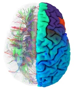 A healthy brain connectome. Image courtesy of Mint Labs/Qmenta.
Our second finalist for Best New Radiology Vendor has developed a cloud-based neuroimaging software platform for hospitals, research centers, and pharmaceutical companies.
Known as Mint Labs in its start-up phase, the company is now in the process of changing its brand name to Qmenta. The Q symbolizes the quality the firm strives for, as well as the quantification it performs on MR images, Mint Labs said. Menta -- derived from the Latin word mensis -- relates to the firm's focus on the brain.
Intended to drive the discovery and development of new treatments for brain disease, the company's advanced visualization and image analytics platform supports data collection, data management, image processing, 3D visualization, and sharing, according to the firm. In May, Mint Labs/Qmenta received a $3 million seed investment, which it plans to use to develop new processing tools to help investigators identify new imaging biomarkers.
Best Radiology Mobile App
CTisus iPearls (iOS), Dr. Elliot Fishman
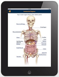 CTisus iPearls. Image courtesy of Dr. Elliot Fishman.
The first finalist for Best Radiology Mobile App comes from a perennial contender for the award. Dr. Elliot Fishman of Johns Hopkins University won for his CTisus Critical Diagnostic Measurements in CT app in 2016, and he was a finalist in 2014 and 2015 for other apps in the CTisus family.
The latest CTisus candidate to vie for Best Radiology Mobile App is iPearls, an iOS app that features "pearls" -- or facts or bits of knowledge -- that can help radiologists make the correct diagnosis or better understand an image. Version 5.0, which was finalized in May, added more than 400 pearls and two new categories.
The app now has thousands of pearls gathered from the literature or the institution's imaging experiences. As is the case with all CTisus apps, iPearls is available for free.
Radiology Assistant 2.0 (iOS), BestApps
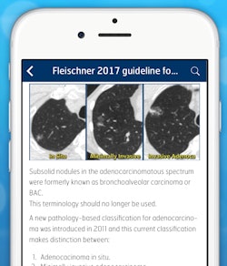 Radiology Assistant 2.0. Image courtesy of Dr. Wouter Veldhuis, PhD.
Developed seven years ago by a Dutch team, Radiology Assistant has been a popular educational app and was even named a finalist for Best Radiology Mobile App in the 2013 Minnies. The latest version has also now achieved finalist status -- just a month after it was released.
Radiology Assistant provides peer-reviewed articles from expert radiologists around the world. With version 2.0, the app was completely rewritten and rebuilt using Apple's new Swift programming language. The result was a significantly improved user experience, including a new article layout and improved searching capability, said developer Dr. Wouter Veldhuis, PhD, a radiologist at University Medical Center Utrecht in the Netherlands. Users can also view inline and full-screen movies and scroll through image stacks, for example.
While Radiology Assistant 2.0 does charge for subscriptions, contributions are used to support the Medical Action Myanmar medical aid organization, according to the developers. Radiology Assistant has been downloaded by users in more than 107 countries.
Previous page | 1 | 2 | 3
