The winners of this year's Minnies awards once again highlight the best that radiology has to offer, with laureates ranging from a radiologist who is head of a major division at the U.S. National Institutes of Health to an app developed as a side project by a U.K. radiographer.
Every year since 2000, AuntMinnie.com has awarded the Minnies, our annual ranking of the best in radiology. Candidates are selected from nominations submitted in August by our members, and winners are chosen through two rounds of voting by our expert panelists. This year's campaign featured 207 candidates in 14 categories, along with the Best Radiology Image winner selected by votes on our Facebook page.
This year's winners offered several surprises. For the first time in years, digital breast tomosynthesis was knocked from its position as Hottest Clinical Procedure. A start-up company less than a year old grabbed the award for Best New Radiology Vendor, while the nod for Scientific Paper of the Year went to a paper that has sparked the interest of the U.S. Food and Drug Administration (FDA) due to its implications for the safety of MRI scans.
Curious about the winners? Check out the profiles below to learn more.
Most Influential Radiology Researcher
Winner: Dr. David Bluemke, PhD, U.S. National Institutes of Health
As director of the Radiology and Imaging Sciences division at the U.S. National Institutes of Health (NIH), Dr. David Bluemke's name has frequently appeared on the list of Minnies candidates, going all the way back to 2005, when he was affiliated with Johns Hopkins University. But 2015 finally marked the year Bluemke won the top spot on the Minnies podium as Most Influential Radiology Researcher.
 Dr. David Bluemke from the U.S. National Institutes of Health.
Bluemke earned both his medical degree and doctorate in biophysics at the University of Chicago, but he spent the next 25 years of his career at Johns Hopkins. While there, he became involved with the Multi-Ethnic Study of Atherosclerosis (MESA), a large population-based study that started in 2000 to track risk factors for cardiovascular disease in a group of 5,000 individuals. Along with Dr. Joao Lima of Johns Hopkins, Bluemke has led the imaging portion of the study, with some two-thirds of the 900 papers published on MESA over the years related to imaging.
Bluemke joined the NIH in 2009, with a particular focus on cardiovascular disease and its complications -- in particular, the use of new imaging technologies to detect subclinical disease. He has been involved in a variety of important research projects over the past year, appearing as a co-author on dozens of research papers, including a number as senior author. Many of these have sprung from MESA.
For example, Bluemke was the senior author on a June 2 paper in Radiology that described how researchers used CT to measure soft plaques and tie their size to risk factors for heart disease. Bluemke and co-authors from NIH and Johns Hopkins found that CT-based measurements of noncalcified plaque corresponded with well-known clinical factors for cardiac disease.
MESA has greatly contributed to a better understanding of the relationship between cardiovascular disease, risk factors, and the structure of the heart. For example, left ventricular mass is now understood to be an important factor in later health outcomes, he believes. Also, it's now recognized that asymptomatic people can have types of carotid artery plaque that put them at high risk of future cardiovascular events.
"These people do not have disease when we look at them, so this gives us the opportunity to intervene early," he told AuntMinnie.com.
In 2015, a new challenge arose for the medical community: Ebola. A number of U.S. healthcare personnel traveled to Africa to help with the outbreak, and several brought the disease back with them -- creating an infection control nightmare for healthcare facilities. The situation dovetailed with another of the NIH's missions: to create public policy that can guide U.S. healthcare practice.
Along with Dr. Carolyn Meltzer of Emory University, Bluemke authored a paper published November 18, 2014, in Radiology that provided recommendations for radiology departments and imaging centers on the proper use of isolation room procedures and protective equipment to prevent infection. They advised that imaging facilities use portable x-ray or ultrasound technologies to image Ebola patients in isolation rooms, rather than risk infecting healthcare staff with the virus while transporting patients to imaging suites -- which could also take hours to disinfect after scanning an Ebola patient.
At NIH's Radiology and Imaging Sciences section, Bluemke oversees 25 radiologists and 200 additional staff. He sees their contribution as essential in being able to perform such influential work.
"The ability to be successful depends on amazing collaborators, not only at NIH but also at Johns Hopkins," he said.
Runner-up: Danny Hughes, PhD, Harvey L. Neiman Health Policy Institute
Most Effective Radiology Educator
Winner: Dr. Geraldine McGinty, Weill Cornell Medicine
The Irish are known for their gift in telling tales, so it's perhaps no surprise that Dr. Geraldine McGinty sees her receipt of the Minnies award for Most Effective Radiology Educator as simply validation of the ability to tell a good story.
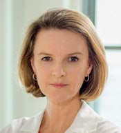 Dr. Geraldine McGinty from Weill Cornell Medical College.
In her case, the subject matter is radiology, and the stories are about how radiologists are adapting to the emerging new environment of value-based care. McGinty has been deeply involved in the area thanks to her activities with the American College of Radiology (ACR) and its Imaging 3.0 initiative.
McGinty was born and raised in the U.K. until the age of 16, when her family moved to Ireland. She received her medical degree in Ireland and completed her residency at the University of Pittsburgh, followed a fellowship in breast imaging at Massachusetts General Hospital.
She'd barely completed the fellowship when she was hired by Montefiore Medical Center to start a new imaging center, which soon grew into an outpatient imaging network. This sparked an interest in practice economics, and Montefiore sponsored her as she completed an MBA at Columbia University in 2000. She then spent 11 years in private practice before joining Weill Cornell Medicine in March 2014.
McGinty said she was inspired to look for ways to give back to the radiology community by Dr. William Thorwarth, who is now CEO of the society. McGinty has been chair of ACR's Commission on Economics for the past four years, and she also works with its Imaging 3.0 project.
McGinty sees the goal of her work at ACR as helping radiologists manage the transition from volume-based payments to value-oriented medicine as mandated by federal initiatives such as the Patient Protection and Affordable Care Act (ACA) and the Medicare Access and CHIP Reauthorization Act (MACRA). MACRA in particular stipulates that by 2022 physicians who are still purely in a fee-for-service environment could see the loss of 9% of their Medicare payments.
While both ACA and MACRA direct radiologists to develop alternative payment models based on value, they haven't provided much guidance on how to do it. That's where ACR and Imaging 3.0 can help, McGinty believes.
The society has been educating its members on the importance of understanding the new initiatives, as well as sharing stories of radiology practices that have adopted value-based payment schemes. It's doing so through vehicles such as webinars, case studies, and presentations at the annual RSNA meeting.
At RSNA 2015, McGinty will participate with other ACR leaders in a session on critical issues facing radiology: in particular, how to transition Imaging 3.0 from a set of talking points into clinical reality. The society sees three major requirements:
- Culture change at radiology practices
- The development of informatics tools to support value-based practice
- The alignment of payment policies with Imaging 3.0
Ultimately, McGinty sees the ability to tell radiology's story effectively as key to the future health of the specialty.
"There is a real sense that we do have to become more patient-focused, we have to tell stories of the value we deliver; otherwise, we won't be recognized for what we do," she said.
Runner-up: Dr. Daniel Kopans, Massachusetts General Hospital
Most Effective Radiologic Technologist Educator
Winner: Chad Hensley, University of Nevada, Las Vegas
Chad Hensley said he got into radiology by accident, but now he wouldn't trade it for anything. He sees the discipline as the intersection through which every other medical specialty passes.
 Chad Hensley from the University of Nevada, Las Vegas.
While attending the University of Nevada, Las Vegas (UNLV) in 1994, Hensley wanted to become a physical therapist. But classes were full, so he tried an x-ray course -- and was hooked.
After completing his training, he worked as a staff radiologic technologist (RT) and then in outpatient imaging for a number of years, but he missed the educational environment and the opportunity to teach. Hensley began teaching a lab in radiologic technology at UNLV's School of Allied Health Sciences, and that soon developed into a full-time staff position in 2003. He's now the clinical coordinator at the program, which has been training technologists for the past 50 years.
One of the projects closest to Hensley's heart is improving the public perception of radiologic technologists beyond the image of "button pushers" that some seem to have of the profession.
"Other medical professionals think they can do our job," Hensley told AuntMinnie.com. "But no one would ever say to a nurse or physical therapist that they could do their job."
Part of the problem is the lack of a clear national standard of minimal education and licensing requirements for technologists. The difficulty in getting such standards passed by the U.S. Congress means that technologists will have to work on a state-by-state basis to develop professional requirements for RTs, Hensley believes.
Ironically, Nevada is one of the states that doesn't require technologists to be licensed. However, Hensley has been working to raise the profile of RTs in the state: In 2014, he helped revive the Nevada Society of Radiologic Technologists and currently serves as its president.
While some newly minted technologists may face a challenging job environment, Hensley believes they have picked a great field -- something he's not afraid to tell his students.
"My priority is to get students to be excited about the field," Hensley said. "They have chosen a profession, they haven't chosen just a job. If they realize the importance that we do, then I'm happy."
Runner-up: Germaine Frosolone, Gateway Community College, New Haven, CT
Most Effective Radiology Administrator/Manager
Winner: Mary Lou Lovel, Wake Radiology
A radiologic technologist by training, Mary Lou Lovel for the past year has been spearheading the migration of a vendor-neutral archive (VNA) at Wake Radiology, a private practice with 50 radiologists and 20 outpatient imaging sites throughout the Research Triangle area of North Carolina.
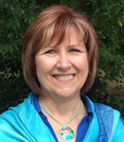 Mary Lou Lovel from Wake Radiology.
Lovel began her career working as a technologist, first at Cape Fear Community Hospital (now part of New Hanover Regional Medical Center) in Wilmington, NC, and then joining Wake Radiology as a CT technologist in 2001. She started acquiring IT experience while at New Hanover when it installed a hospital information system, and when Wake began installing its first RIS and PACS, she found herself on the team that led the transition. This work eventually turned into a formal role as a systems analyst, and Lovel received her certified imaging informatics professional (CIIP) credential four years ago.
Over time, staff at Wake Radiology began to feel that its PACS network, based on the traditional model of a single vendor supplier and proprietary software, wasn't changing with the times. The practice felt that the PACS lacked flexibility -- for example, when another site began sending images to Wake in an uncompressed JPEG 2000 format, Wake's PACS wasn't able to accept them.
What's more, images were archived in a format proprietary to the PACS vendor, and Wake's IT staff had to "jump through hoops" to make any changes to DICOM data. Wake realized it was time for a change.
"We knew we needed a VNA because we didn't want proprietary images to be stored again, and because we didn't want to be reliant on the vendor," she said.
Lovel and Wake's clinical systems manager, Brian Appleby, were intrigued by the concept of a "deconstructed PACS," which departs from the traditional approach based on a single vendor. Instead, it makes the VNA the center of operations, with other elements such as viewers, reporting modules, and workflow engines added with a best-of-breed approach.
They began implementing a VNA at the beginning of 2015, starting with Wake's mammography service, which recently adopted digital breast tomosynthesis (DBT). Mammography had presented a particular challenge for Wake, which used a batch reading protocol and dedicated mammography workstations. Under the old system, images were being sent three to four times across the local area network to reach different workstations. With the new architecture, Wake now has a workflow engine that routes studies to the correct workstation based on study description, reducing the amount of time from what used to be required to manually push mammography studies.
Lovel described Wake's experiences in a presentation at the Society for Imaging Informatics in Medicine (SIIM) meeting in May. Meanwhile, Wake's data migration to the VNA is still ongoing, with the site currently working on implementing viewer software.
What's Lovel's advice for sites thinking about the VNA route? She advises them to have the VNA in place first before adding other elements like the viewer.
"Get your migration into a VNA done before you start finding a new viewer and replacing a lot of parts at the same time," she said. "The migration is the hardest part -- get that done before you start doing anything else. When you have a lot of moving parts, it's hard to keep the end users understanding what's happening."
Runner-up: Charles Powell, Emory University
