A new PET imaging technique called C-11-PABA PET accurately detects and monitors Mycobacteroides abscessus lung infections, according to research presented June 24 at the Society of Nuclear Medicine and Molecular Imaging (SNMMI) meeting.
These infections are some of the most difficult-to-diagnose in patients with chronic lung diseases, and the technique could offer a new diagnostic tool that would improve diagnosis and treatment, said lead study author Yuderleys Masias-Leon, MD, of Johns Hopkins University Medical School in Baltimore, MD, in an SNMMI statement.
"Given the constraints of conventional imaging, the development of noninvasive, bacteria-specific imaging techniques would be clinically valuable for diagnosing and selecting appropriate treatments for patients," she said.
Mycobacteroides abscessus is a fast-growing mycobacterium that affects immunocompromised patients and those with underlying lung diseases, such as cystic fibrosis or chronic obstructive pulmonary disease, Masias-Leon and colleagues explained. Conventional imaging tends to be limited in distinguishing the bacterial infection from these inflammatory diseases -- and "definitive diagnosis often requires invasive procedures," they wrote.
The investigators tested the C-11-PABA PET technique in both preclinical and clinical settings; in vitro studies confirmed that the tracer accumulated in the bacteria. They then performed C-11-PABA PET exams with mice before imaging a 33-year-old patient with cystic fibrosis and microbiologically-confirmed Mycobacteroides abscessus pulmonary infection.
The group found the following:
- C-11-PABA uptake was observed in all in vitro studies.
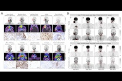
- Sustained accumulation of the tracer was found in the lung lesions of Mycobacteroides abscessus-infected mice.
- In the patient with cystic fibrosis, C-11-PABA PET showed significantly higher PET uptake in the affected lesions compared to unaffected lung -- which confirmed the pulmonary bacterial infection.
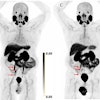
 Representative coronal (left) and transverse (right) CT and C-11-PABA PET/CT images from a 33-year-old female with cystic fibrosis and confirmed M. abscessus infection in a 45-minute dynamic PET scan after an intravenous injection of 518 MBq of C-11-PABA. In the first nine minutes of the scan C-11-PABA got distributed systemically, followed by rapid clearance via heptic and renal excretion, showing accumulation of C-11-PABA only inside the affected lesion in the last 10 minutes. Not all lesions visualized on CT were PET avid, suggesting that some lesions may be sterile or had very low bacterial burden. The y axis shows time in minutes. Image and caption courtesy of the SNMMI and Yuderleys Masias-Leon, MD.
"This proof-of-concept data highlights the potential of C-11-PABA PET as an innovative, bacteria-specific, noninvasive and clinically-translatable diagnostic tool," Masias-Leon said. "In the future, C-11-PABA PET could offer rapid and accurate diagnosis to facilitate precision medicine and individualized care for patients with this infection."