PET/CT imaging times can be reduced significantly for women with suspected breast cancer, according to a study published December 8 in Scientific Reports.
The finding is from a group in China who were able to differentiate between malignant and benign lesions on PET/CT images acquired at both 10 and 60 minutes after F-18 FAPI-04 radiotracer injection.
“[Early imaging] is not only more patient-friendly but also has the potential to simplify clinical workflows and reduce unnecessary biopsies,” noted lead author Zonglin Li, MD, of The Second Affiliated Hospital of Guangxi Medical University in Nanning, and colleagues.
Molecular imaging agents based on fibroblast activation protein inhibitors (FAPI) have emerged as a promising tool in breast cancer diagnosis, as fibroblast activation protein is highly expressed in the tumor microenvironment of the disease, the authors explained.
However, most existing studies have focused on late imaging performed approximately 60 minutes after tracer injection. Such prolonged waiting may be impractical for patients unable to tolerate the delay, they noted.
To investigate the feasibility of using early F-18 FAPI-04 PET/CT, the group recruited 40 women with suspected breast cancer (mean age, 52) and performed dual-phase PET/CT (Biograph Sensation 16, Siemens Healthineers) at approximately 10 minutes (early) and 60 minutes (late) after injection of F-18 FAPI-04 radiotracer.
Prior to PET/CT imaging, none of the patients had received any treatment, including surgery, chemotherapy, radiotherapy, or immunotherapy. Histopathological diagnoses were obtained through biopsy or surgical resection, both performed after PET/CT imaging.
Two nuclear medicine experts with more than 10 years of experience in nuclear medicine diagnostics interpreted all PET/CT images independently, with any discrepancies in interpretation resolved by consensus-based review.
According to the analysis, a total of 51 breast lesions were detected, with the researchers obtaining maximum standard radiotracer uptake values (SUVmax) by outlining regions of interest in the images. After the PET/CT scans, all lesions underwent tissue biopsy, with 36 being malignant breast cancer and 15 being benign breast lesions.
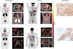
 The characteristic imaging features of high-risk breast lesions on early and late F-18 FAPI-04 PET/CT. (A, D) are CT images; (B, E) are early imaging fused images; (C, F) are late imaging fused images. (A–C) A 35-year-old patient diagnosed with invasive ductal carcinoma, as indicated by the red arrow, with ultrasound findings classified as BI-RADS 4a. In the early and late imaging, the maximum standard uptake value (SUVmax) (12.5 vs. 14.2), lesion-to-background ratio (LBR) (22.2 vs. 22.2), respectively. (D–F) A 51-year-old patient with a breast fibroadenoma, as indicated by the white arrow, also categorized as BI-RADS 4a. In the early and late imaging, the SUVmax (0.5 vs. 1.1), LBR (2.2 vs. 2.6), respectively. These findings demonstrate that both SUVmax and LBR in breast cancer are markedly higher than those in benign breast lesions across both imaging phases.Scientific Reports
At these cutoff values, early imaging with SUVmax achieved the highest diagnostic performance, with an AUC of 1, and both sensitivity and specificity of 100%. There was no significant difference in diagnostic performance between early and late imaging, the researchers reported.
“Our findings indicate that early F-18 FAPI-04 PET/CT imaging offers diagnostic accuracy comparable to late imaging while significantly reducing acquisition time, supporting its potential as a practical alternative in clinical practice,” the group wrote.
To more comprehensively assess the clinical value of F-18 FAPI-04 PET/CT in the diagnosis of breast lesions, future studies should include suspected breast cancer cases from multiple medical centers, the researchers noted.
“Moreover, further investigations are warranted to explore the potential applications of this technique in breast disease subtyping, staging, and treatment response evaluation,” the group concluded.
The full study is available here.