PET imaging improves oligometastatic breast cancer detection over conventional imaging, according to a post-hoc analysis presented June 21 at the Society of Nuclear Medicine and Molecular Imaging (SNMMI) meeting in New Orleans, LA.
In his presentation, Ur Metser, MD, from the University of Toronto in Ontario, Canada, discussed his team’s findings on how PET imaging finds oligometastatic disease (OMD) in women 2.5 times more often than conventional imaging. And PET detects more extensive locoregional metastatic disease, the team added.
“These discrepancies in performance may impact patient selection for metastasis-directed therapy,” Metser said.
It remains unknown how women with oligometastatic breast cancer can be best managed, with researchers continuing to research methods for selecting women who may benefit from metastasis-directed therapies.
Oligometastatic breast cancer is considered an intermediate stage between localized and widely metastatic disease, where the cancer has spread to locations outside the breast and nearby lymph nodes. The number of locations falls between one and five.
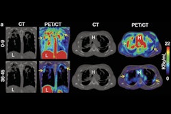
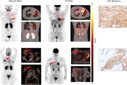
Metser and colleagues investigated the prevalence of OMD in women with locally advanced breast cancer at initial staging with F-18 FDG PET/CT or conventional imaging. They also compared patterns of locoregional and distant metastatic disease.
The team performed its post-hoc analysis from its own prospective, multicenter randomized trial consisting of 369 women with stage IIb (T3N0) or III invasive breast cancer. The women were randomized to staging with PET (n = 180) or conventional imaging consisting of CT chest, abdomen, and pelvis exams and bone scintigraphy (n = 185). The previous study found that the conventional approach underestimates disease extent, which may worsen proper patient selection for ablative therapies.
The PET approach in the current study detected OMD in 19 women (10.6%) compared with eight found through the conventional approach (4.3%, p = 0.02). PET also found extensive metastatic disease (> 5 distant metastases) in 24 women (13.3%) versus 13 found on conventional imaging (7.1%, p = 0.04).
All women with OMD on PET and conventional imaging had axillary lymph node metastases. However, PET found extra-axillary regional lymphadenopathy and extra-regional lymph node and liver metastases more often.
Frequency of imaging findings detected on PET, conventional imaging | ||
Finding | Conventional | PET |
Extra-axillary regional lymphadenopathy | 12.5% | 31.6% |
Extra-regional lymph node | 0% | 15.8% |
Liver metastases | 12.5% | 31.6% |
Metser reiterated that these findings may help optimize appropriate patient selection for metastasis-directed therapy.
“Based on retrospective data suggesting long-term, disease-free intervals, the European Society of Medical Oncology [ESMO] recommend considering multimodal therapy with curative intent in patients with oligometastatic breast cancer, who are amenable to ablative therapy,” Metser added. “But recent data are not as encouraging.”
He also called for more randomized trials in the future to find out whether these therapies for PET-defined oligometastatic breast cancer improve outcomes.