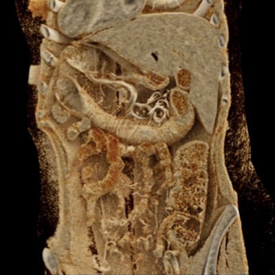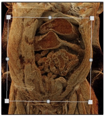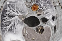
The photorealistic quality of cinematically rendered CT scans can aid in the understanding of complex colon anatomy and has the potential to facilitate the detection and characterization of colon pathology in three major ways, according to an article published in the May/June issue of the Journal of Computer Assisted Tomography.
Though still in its infancy, cinematic rendering has already shown promise in a variety of clinical applications, from evaluating spleen anatomy and pelvic bone to aiding diagnosis in aortic complications. The technique relies on a global lighting model to produce photorealistic images with high surface detail from any volumetric CT dataset; it takes an experienced radiologist roughly five minutes to create these images using computer software (syngo.via VB30, Siemens Healthineers).
"At our institution, cinematically rendered images are routinely generated in many situations including initial diagnosis and follow-up of a variety of malignant conditions, before and after surgical and minimally invasive vascular procedures, and after trauma," wrote a team from Johns Hopkins University led by Dr. Steven Rowe, PhD (J Comput Assist Tomogr, May/June 2019, Vol. 43:3, pp. 475-484).
 Cinematically rendered CT scan of the colon in patients with Crohn's disease (top) and ulcerative colitis (bottom). All images courtesy of Dr. Elliot Fishman.
In the article, the researchers discussed the potential role that cinematic rendering could have in the evaluation of different types of colonic pathology:
- Inflammatory conditions: Colitis often presents with a wide range of symptoms, making it relatively difficult for radiologists to pinpoint underlying causes and detect potential complications on standard imaging alone. Cinematic rendering offers a highly detailed view of the surface of high-density structures, such as vessels, as well as low-density soft tissues that could be even more useful than 3D CT in providing supplemental visualization, the authors noted.
- Neoplastic pathology: For the evaluation of colorectal cancer, CT scans can facilitate the detection of metastatic disease and the delineation of tumor invasion and staging. However, standard CT is limited in its capacity to show disease-involved lymph nodes and determine the depth of tumor invasion. Cinematic rendering offers a much higher level of surface detail of textural features than standard CT, enabling the 3D technique to contribute to tumor characterization as well as surgical planning, the authors noted. Clinicians can also vary window width and level settings on cinematically rendered images to improve the distinction between tumor tissue and neighboring structures even more than with traditional volume rendering and maximum intensity projection (MIP).
- Hernias and bleeding: Cinematically rendered CT angiography scans may aid the examination of inguinal hernias and lower gastrointestinal bleeding. The technique can effectively display the spatial arrangement of blood vessels along the colon wall as well as capture contrast extravasation in patients who are actively bleeding. Combining the utility of traditional volume rendering and MIP protocols, cinematic rendering allows for the identification of soft-tissue abnormalities while maintaining high enough contrast to reveal subtle extravasation.
Diverticulitis is another common inflammatory condition evaluated on CT scans. Though cinematic rendering is unlikely to help radiologists spot key features of diverticulitis better than traditional CT, the photorealism of cinematically rendered images may prove valuable in preoperative planning for cases requiring surgical intervention. In one case at the group's hospital, clinicians were able to spot an inflammatory pseudopolyp in a patient using cinematic rendering and then appropriately manage it before resecting the colon.
"Indeed, a great deal of work remains to be done to validate the utility of cinematically rendered images in clinical practice, although the technique is promising given the enhanced detail and realistic shadowing," Rowe and colleagues wrote.
If early case studies are any indication, cinematic rendering can be useful for the preoperative assessment of colonic disease and response to therapy as well as the correlation of findings, they added. What's more, the technique will almost certainly be applied in engaging patients and educating trainees.


