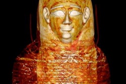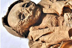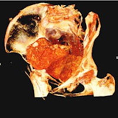
The patient presented after 3,000 years. X-ray and CT scans revealed first that this was not a male scribe and priest, as suspected, but in fact a woman about 25 years old, possibly a society elite from a vast necropolis in Thebes in Upper Egypt.
Moreover, the woman was pregnant when she was mummified.
The find is the only known case of an embalmed pregnant woman, according to Polish researchers from the National Museum in Warsaw, who published results of the investigation April 28 in the Journal of Archeological Science. The discovery opens possibilities for a new understanding of pregnancy in ancient times, the development of fetuses, and the taphonomic processes of fetuses as well as uteruses.
"Our first surprise was that it has no penis, but instead it has breasts and long hair, and then we found out that it's a pregnant woman. When we saw the little foot and then the little hand, we were really shocked," said Marzena Ozarek-Szilke, an archeologist at the University of Warsaw, in a statement to the Associated Press.
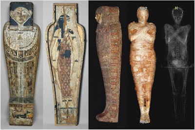 The coffin, cartonnage case, and mummy. All images courtesy of the Journal of Archaeological Science.
The mummy, its coffin, and its cartonnage case have been among a collection of ancient artifacts in the National Museum in Warsaw since about 1917. The object was acquired in Egypt and donated to the University of Warsaw in December 1826. The craftsmanship and style of the coffin date the object to about the 1st century B.C. According to the inscription on the assemblage, the mummified Egyptian was a male scribe and priest.
The museum investigators enlisted the help of a radiology team from a local Affidea Clinic in Otwock that performed the imaging using a portable x-ray unit (Optima XR220amx, GE Healthcare) and CT scanner (CT Big Bore, Philips). As a result, the following new information regarding the content of the bundle was obtained:
- Fractures of the ethmoid bone showed that the brain was removed through the nasal cavity and that the cranial cavity was filled with an embalming substance.
- Four mummy-shaped amulets with animal or human heads measuring approximately 4 cm each were seen in the chest of the mummy. These so-called "four sons of Horus" are divinities protecting the woman's viscera. At least 15 objects may have been included among the wrappings of the mummy.
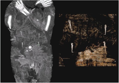 The abdominal area of the mummy with amulets representing the four sons of Horus above the navel area.
- The sex of the mummy was confirmed by the fetus, breasts, and female genitalia seen on the CT images. Her teeth are well preserved and slightly worn, with the tips of the roots of the third molars completely formed. The squamosal suture is not obliterated. From this, the researchers determined the woman died between the ages of 20 and 30 years old.
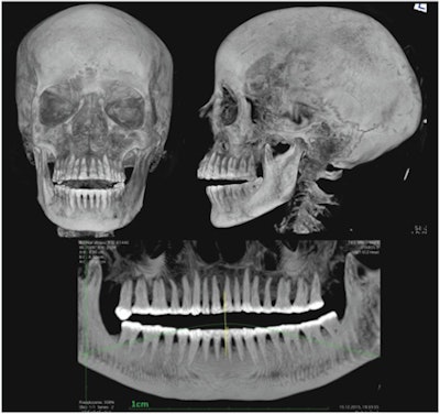 X-ray of the head and the pantomographic image of the teeth of the mummy.
- The fetus was mummified together with its mother. Its head circumference is 25 cm, which indicates that it was between 26 and 30 weeks old. The poor state of the child's skeleton made it impossible to take any measurements of other bones, the researchers wrote.
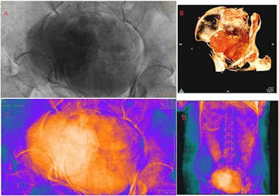 Abdominal area of the mummy: (A) x-ray; (B-D) CT.
The woman was carefully mummified, wrapped in fabrics, and equipped with a rich set of amulets. Above all, the researchers wrote, it is unknown who she was. They did name her, however: "The Mysterious Lady of the National Museum in Warsaw."
The collection of ancient Egyptian mummies preserved at the National Museum in Warsaw has rarely been studied in detail. This research was part of the Warsaw Mummy Project, which was launched in 2015 by the Polish Academy of Sciences with the aim of conducting a comprehensive analysis of all human and animal mummies at the museum.
Further studies of this specimen and others may prove to be beneficial for medical science, especially obstetrics and radiology, the researchers stated.
"The fact that only noninvasive examinations of this mummy have been conducted so far means that it is intact and can be the subject of future multidisciplinary investigations," they concluded.


