Dear Advanced Visualization Insider,
Vertebral compression fractures are a serious problem, affecting approximately 750,000 people annually and as many as one-fourth of all postmenopausal women. They can be as debilitating for patients as they are difficult for clinicians to diagnose.
But researchers from the U.S. National Institutes of Health are taking aim at that last problem with a new computer-aided detection (CAD) scheme. Their approach speeds up the process of diagnosing vertebral compression fractures with high sensitivity and a very low false-positive rate.
The algorithm features a novel "height compass" that measures the height and axial plane angle of each vertebra, looking for signs of fracture. It works very well, as you'll learn in our Insider Exclusive.
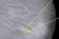
Two CADs are better than one in digital breast tomosynthesis (DBT), according to a study from the University of Michigan. The study group's technique applies two independent CAD algorithms in the search for microcalcification clusters: first to data acquired with a new low-dose DBT protocol, then to planar projection images derived from DBT data.
Using two algorithms allowed the researchers to halve the dose while maintaining impressive results. Get the rest of the story by clicking here.
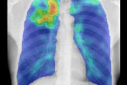
Moving to the lungs, radiography still has a role in tuberculosis diagnosis, but owing to wide differences in exposure and postprocessing by different digital radiography (DR) systems, it's difficult to apply automated analysis to the images and get anything useful out of them.
However, during a presentation at the RSNA 2014 meeting, Dutch researchers explained how they developed a tool for "normalization" of the DR images, which enabled CAD to distinguish likely cases of tuberculosis. Find out how well it works by clicking here.
Stay tuned for more news in your Advanced Visualization Community, and be sure to scroll through the rest of the links below.


