Dear AuntMinnie Member,
What does renowned animation studio Pixar have to do with radiology? Plenty, according to Pixar's president, Ed Catmull, PhD, who addressed the topic in an article published last week in the Journal of the American College of Radiology.
Even though Pixar has been fabulously successful over the years, it has made plenty of mistakes on its way to creating cinematic magic. But Pixar learned from its missteps and created a corporate culture based on candor and risk-taking that has helped it maintain its creative edge.
Learn about how these lessons can apply to medical imaging -- and why Mr. Catmull was speaking about radiology in the first place -- by clicking here for an article in our Imaging Leaders Community.
CAD for vertebral fractures
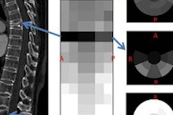
In other news, a team from the U.S. National Institutes of Health (NIH) recently presented a computer-aided detection (CAD) algorithm that it developed to detect vertebral compression fractures on spine CT images.
The NIH team led by Dr. Ronald Summers, PhD, developed a quantitative system for fractures that measures minute changes in the height of vertebrae. Learn more by clicking here, or visit our Advanced Visualization Community at av.auntminnie.com.
Reducing DSA dose
Finally, the Society of Interventional Radiology meeting starts on Saturday in Atlanta, and we're pleased to bring you a sneak peek at a Sunday presentation in which researchers from Cincinnati Children's Hospital Medical Center will discuss their efforts to reduce radiation dose in pediatric digital subtraction angiography (DSA) studies.
The group started with a commercially available protocol for reducing dose in adult DSA studies, and tweaked its technical parameters to wring out even greater reductions for pediatric exams. In fact, they have achieved a 95% reduction in dose compared to conventional DSA -- while still producing diagnostic-quality images.
Learn more by clicking here, or visit our Digital X-Ray Community at xray.auntminnie.com.
