Dear Advanced Visualization Insider,
Computer-aided detection (CAD) in radiology has nearly always been trained to find abnormalities in medical imaging data. Until now, that is. A new CAD technique -- which might more properly be called computer-aided diagnosis -- goes a step further by generating radiology reports complete with differential diagnoses based on imaging findings.
Developed at Oregon Health and Science University and presented at the Society for Imaging Informatics in Medicine (SIIM) meeting last week, the new technique pores through imaging findings and generates -- on a good day, at least -- quite plausible lists of differential diagnoses at lightning speed. The tool, based on readily available automated data sources that are constantly improving, has many promising uses, according to its developers. Get the rest of the story in this issue's Insider Exclusive.
In a similar vein, CAD is also being recruited to help researchers pluck select cases from a sea of patient data stored in hospital PACS networks, only a few of which might have the particular set of subtle findings that qualify them for a research project.
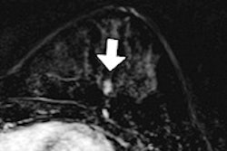
Researchers from Detroit's Henry Ford Hospital decided to tackle what was becoming a major time waster: finding patients who qualify for research studies from a PACS loaded with nearly a million cases. The algorithm sorted through all of it much faster than humans could, generating about 700 knee MRI cases that were perfect for their research. Potential uses for the hybrid data-mining/CAD application are many and varied, say the researchers in an article you'll find here.
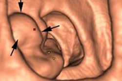
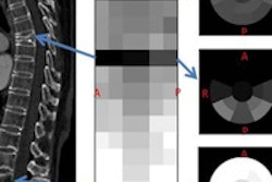
Speaking of time savers, yet another algorithm has been developed to semiautomatically segment a wide array of brain tumors, offering radiologists a one-stop shop for patients with multiple lesions. Based on an adaptive geodesic algorithm, the new technique is fast and accurate, according to this story.
Also from the advanced visualization and CAD sessions at SIIM 2015, a new technique from Emory University targets dangerous medical errors head-on by putting patient photos right in the DICOM archive, next to the imaging study.
Before they rolled out the new functionality, the investigators wanted to know what various physicians and patients might think about this new potential intrusion on patient privacy. Only one group was surprisingly wary of the idea -- and not due to privacy concerns. Find out who it was by clicking here.
For those who want to go really big, an Israeli start-up promises to crunch vast numbers of patient datasets for use by industrial-scale firms like healthcare companies that want to look at disease trends in populations, among other potential applications.
Click here for the story, or visit your Advanced Visualization Community to stay up to date with the burgeoning world of automated applications for radiology.