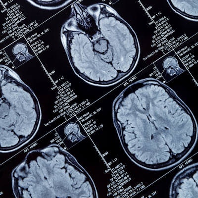
Radiologists who used structured reporting to interpret MRI scans of glioma patients produced reports that were judged to be more consistent and easier to understand than conventional free-text reports, researchers found in a study published online August 27 in Academic Radiology.
A group led by Dr. James Zhang, PhD, from Emory University analyzed quantifiable metrics in post-treatment MRI reports for glioma patients prepared before and after adoption of the Brain Tumor Reporting and Data System (BT-RADS), which was previously developed at Emory. Zhang and colleagues found quantifiable and objective benefits from the use of BT-RADS.
"As compared to conventional free-prose reports, reports derived from BT-RADS demonstrated superior traits across multiple domains, including greater consistency and completeness of history reporting, reduced ambiguity, and improved conciseness," they wrote.
A multidisciplinary group at Emory had previously developed BT-RADS with the goal of improving the classification and reporting of post-treatment brain tumor images, with a particular focus on primary gliomas, which are difficult to report consistently, according to the authors.
"Because of the subjectivity of conventional free-prose reporting, this can introduce variability, inconsistency, and ultimately confusion in reporting," the authors wrote. "This can lead to miscommunication between the radiologist and other members of the healthcare team, and even between different radiologists who may be interpreting the same images at varying time points."
A preliminary, subjective survey at their institution found that BT-RADS had yielded improvement in three previously identified shortcomings associated with free-prose reports: insufficient clarity in communication, inadequate consistency in reporting, and too much ambiguity. Next, the researchers sought to determine if there were also objective benefits to BT-RADS.
To accomplish this, they analyzed discrete, quantifiable metrics, including the use of history words such as "Avastin" and "methylguanine-DNA methyltransferase" and hedge words such as "possibly" and "likely." They performed this analysis on post-treatment glioma MRI reports from three-month periods before (211 reports) and after (172 reports) the adoption of BT-RADS. The reports were written by six different board-certified and fellowship-trained neuroradiologists.
Zhang and colleagues found a significant improvement in all metrics from the use of BT-RADS.
| Quantifiable metrics in post-treatment MRI reports for glioma | |||
| Reporting metric | Free-prose reporting | BT-RADS | p-value |
| Use of "Avastin" | 7.6% | 20.9% | < 0.001 |
| Use of "methylguanine-DNA methyltransferase" | 10.9% | 31.4% | < 0.0001 |
| Use of hedge word "possibly" | 3.8% | 0.6% | < 0.05 |
| Use of hedge word "likely" | 49.8% | 28.5% | < 0.01 |
| Mean length of impression section | 53.7 words | 42.6 words | < 0.01 |
| Mean total report length | 389 words | 245.2 words | < 0.001 |
| Reports including addenda | 10% | 1.2% | < 0.01 |
"Given these findings, BT-RADS is a promising new structured report system that can benefit any institution performing imaging following glioma treatments," they wrote. "Furthermore, these results support further investigation into additional fields that could be supplemented with structured reports."
The researchers are currently conducting ongoing studies investigating reader agreement and concordance with tumor board decisions. These future investigations may further corroborate these findings and help optimize the scoring parameters, according to the group.


