Healthcare disparities continue to plague medical imaging, but there are concrete measures radiologists can take to mitigate them, according to a paper published on October 12 in RadioGraphics.
"Radiologists and radiology practices can become active partners in efforts to assist patients along their imaging journey and overcome existing barriers to equitable cancer screening care for traditionally marginalized populations," wrote a group led by Peter Abraham, MD, of the University of California, San Diego.
Avoidable differences in disease burden and outcomes that socially disadvantaged individuals may experience manifest in nearly all areas of radiology, from emergency imaging, neuroradiology, and nuclear medicine to image-guided interventions and imaging-based cancer screening, the group noted. These inequalities can have substantial effect on population health, it explained.
"Disparities in imaging-based cancer screening, especially breast cancer screening and breast cancer outcomes, are currently the most well-documented health disparities in radiology and are especially noteworthy given the far-reaching population health impact," the group wrote.
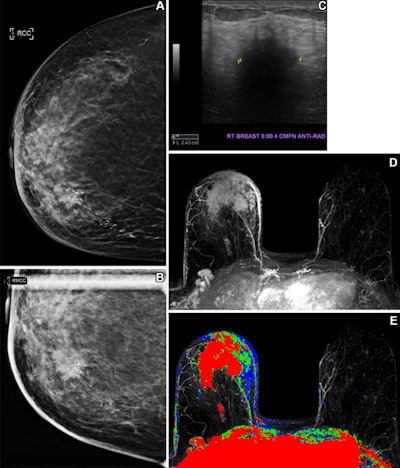
 Health insurance coverage influences breast cancer screening behavior. In one case, a 61-year-old uninsured woman with no known previous screening for breast cancer presented for screening mammography. (A) A craniocaudal two-dimensional synthetic mammogram of the right breast shows an irregular mass in the medial breast with pleomorphic calcifications extending posteriorly. (B) A craniocaudal magnified mammogram more clearly shows the
irregular mass and pleomorphic calcifications in the medial breast. (C) An anti-radial ultrasound image of the right breast shows an irregular hypoechoic mass with indistinct margins and posterior acoustic shadowing at the 3 o'clock position, approximately 4 cm from the nipple. (D) An axial postcontrast maximum intensity projection (MIP) subtracted MR image shows a large enhancing mass in the anterior and medial right breast with skin thickening and enhancement and several morphologically abnormal right axillary lymph nodes. (E) An axial postcontrast MIP subtracted MR image with color mapping shows the rapidly enhancing mass with washout kinetic enhancement in the anterior and medial right breast, with skin thickening and enhancement and several morphologically abnormal right axillary lymph nodes. Histopathologic biopsy results demonstrated invasive mammary carcinoma with metastatic disease in a right axillary lymph node. Image courtesy of Peter Abraham, MD, et al.
But radiologists have a significant role to play in addressing the problem, since the specialty "exists at the intersection of diagnostic imaging, image-guided diagnostic intervention, and image-guided treatment," the authors explained.
The team outlined seven ways to reduce healthcare inequalities in imaging, ranging from engaging in the wider political arena to offering patients practical support:
- Advocate for a supportive health policy.
- Work to reduce patients' out-of-pocket costs.
- Lobby for increased price transparency.
- Improve education and outreach efforts for screening exams.
- Ensure that appropriate language translation services are available in the radiology department.
- Offer individualized assistance with appointment scheduling.
- Make sure transportation assistance and childcare are available.
Radiologists "have an imperative to familiarize themselves with the role that social determinants of health play in creating disparities in radiologic care and health outcomes," the authors wrote.
"Through improved understanding of how social determinants of health can drive differences in health outcomes in radiology, radiologists can effectively provide patient-centered, high-quality, and equitable care," they concluded.
The complete study can be found here.


