VIENNA - What do an egg, a pair of scissors, a screwdriver, and buttons have in common? ECR delegates puzzled over the connection before Dr. José Cáceres provided the answer: They haven't changed since their creation. They are the perfect design.
The chest x-ray has been similarly enduring. While CT has undergone three generations of design in only 27 years, the general way chest x-rays are performed and appear has not altered in 115 years. In Friday's Josef Lissner Honorary Lecture, Cáceres explained that if the original radiologists from the turn of the 20th century were transported to the present day, they would have little difficulty performing a modern chest radiograph.
With the exception of Marilyn Monroe's chest x-ray, sold last year to a private collector for $45,000, plain film is relatively low cost, at 15 times less than the cost of a CT exam, and 80 times lower than a low-dose CT -- and it is speedily obtained. Radiologists also tend to interpret images quickly, he added, meaning that chest x-rays were ubiquitous in today's clinical setting, their ratio vastly outnumbering that of CT scans across most of Spain's hospitals.
This speed, however, could prove to be a double-edged sword for the patient. If x-rays were not conclusive, some doctors would simply repeat a chest image with contrast or opt for a CT, according to Cáceres, chief of diagnostic radiology at HGU Vall d'Hébron, Barcelona. More careful scrutiny of chest radiographs was key to better diagnosis.
"My own personal formula is I = bk + lv + pf x e, in other words, Information equals basic knowledge plus lateral view plus previous films ... all multiplied by enthusiasm," he said.
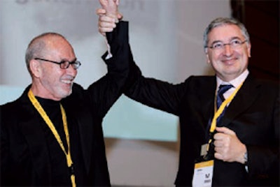
 Bravo, mon ami! ECR Congress President Dr. Yves Menu warmly congratulates Dr. José Cáceres after his honorary lecture.
Radiologists should bear in mind three commandments. The first, thou shalt not forget basic knowledge, involves knowing conditions such as hypogenetic lung, which will facilitate plain film diagnosis without the need for CT.
The second, thou shalt not forget the lateral view, hinges on the fact in 1994 Chotas et al estimated that 26.4% of lung volume was obscured by cardiac mediastinal and subdiaphragmatic structures. Lateral views complementing conventional views can pick up metastases earlier.
The third commandment, thou shalt not forget to look at previous studies, can help radiologists to pinpoint existing pathologies that could otherwise eat up time and resources. A case in point was a strange shadow on the heart, imaged on a patient in August 2010 at Cáceres' hospital. The shadow was missed in CT owing to it being the same opacity as the contrast used. Previous plain film images from November 2007 showed the same shadow, the diagnosis proving to be casseous calcification of the mitral annulus.
"To detect disease and determine the next step, chest x-ray complements CT, not competes with it," he said.
Before he was presented with the ECR's honorary diploma, Cáceres highlighted how the accumulative experience gained over a century of chest x-ray was today's radiologists' inheritance. For the individual doctor, chest x-ray "could not be learned quickly" and might even take a lifetime.
Originally published in ECR Today March 5, 2011.
Copyright © 2011 European Society of Radiology


