Certain advanced MRI metrics are more useful than others for assessing white matter alterations that could be linked to long-COVID brain fog, according to research results presented May 6 at the International Society for Magnetic Resonance in Medicine (ISMRM) meeting.
A team led by Nicolò Rolandi, PhD, of University College London in the U.K. explored voxel-wise analysis of core white-matter voxels using advanced MRI metrics and neuropsychological scores.
The study dataset included 25 healthy control participants and 47 with long COVID who were scanned and clinically assessed between June 2020 and August 2023. MRI data were acquired using a Philips Ingenia CX 3T scanner with the product 32-channel head coil, the group noted.
The investigators used quantitative magnetization transfer, inversion recovery, and diffusion-weighted imaging to calculate advanced MRI metrics. They assessed changes of bound pool fraction (BPF), spin-spin relaxation time of the bound pool (T2B), longitudinal relaxation time (T1), fractional anisotropy (FA), mean diffusivity (MD), orientation dispersion index (ODI), and neurite density index (NDI).
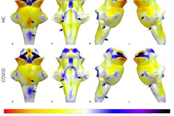
Each showed regional microstructural alterations at a voxel-wise level. The team noted that voxel-wise analysis of the core skeleton of white matter used 5,000 permutations and corrected for multiple comparisons using threshold-free cluster enhancement (TFCE).
The insights generated could add to the medical community's understanding of the involvement of white matter alterations as either a risk factor or consequence of long COVID. For ISMRM, the team noted four interesting findings:
- T2B widespread alterations were observed in the frontal lobe, diencephalon, and left occipital lobe, with increased T2B values being observed in long COVID compared with healthy controls.
- Widespread alterations of BPF were found in the frontal lobe and more localized to the left temporal and occipital lobes, with reduced BPF being observed in long COVID.
- No alterations were found using FA, MD, ODI, and NDI.
- No correlation was found between SDMT and MRI metrics.
This research adds to previous studies that have shown brain structural alterations following COVID-19. It is important because it highlights possible mechanisms associated with brain fog in people experiencing long COVID.
Approximately 30% of people who develop COVID after contracting SARS-CoV-2 experience a variety of symptoms that after 12 weeks, are defined as long COVID or post-COVID-19 syndrome, the authors noted. Lingering issues may include neurological symptoms, such as trouble thinking and concentrating, that are a type of brain fog.
Using MRI would enable a more thorough exploration of the diverse cognitive dimensions affected in people experiencing brain fog and perhaps a better understanding of how white matter alterations are involved, Rolandi and colleagues concluded.