HONOLULU - Synthetically generated "postcontrast" screening breast MR images show promise as an alternative to contrast MRI exams in women with contrast-related contraindications, according to research presented May 12 at the International Society for Magnetic Resonance in Medicine (ISMRM) meeting.
The findings could make MR imaging for breast cancer screening more available, wrote a team led by Su Min Ha, PhD, of Seoul National University Hospital in South Korea.
"There is a growing interest in noncontrast breast MRI to make breast cancer detection more accessible, for patients at risk from gadolinium contrast agents," the authors explained. "Synthetic postcontrast MRI may broaden MRI applications for diagnostic and screening purposes."
Breast MRI is considered to be the most sensitive imaging tool for breast cancer detection, but concerns about gadolinium retention and access issues limit its use, the team noted. Ha and colleagues investigated the use of a deep-learning method to generate synthetic "postcontrast" breast MR images from noncontrast exams for breast cancer detection. They used an Extra-Dimensional U-NET algorithm with visual geometry to simulate T1-weighted postcontrast images from precontrast T1-weighted and diffusion-weighted images (DWI), and compared the sensitivity of synthetic postcontrast breast MR images, noncontrast MR images (including T1-weighted and diffusion-weighted imaging), and the combination of the two.
The study consisted of 279 breast cancer patients with 282 lesions identified between June 2019 and December 2020. The group used 500 MRI exams culled from this cohort to train the algorithm, and two breast radiologists assessed cancer detectability on a 5-point scale (with 5 equal to excellent and 1 equal to unacceptable).
The researchers found the following:
- Breast cancer detectability on synthetic MRI reached 89% on average by the two readers, and 91.8% on noncontrast MRI (p = 0.006).
- Combined synthetic and noncontrast MRI increased breast cancer detection sensitivity to 95.4%, outperforming each method alone (p < 0.001).
- Lesions identified only by synthetic MRI were primarily nonmass lesions (55%), smaller than 1 cm (4%), and mucinous cancer (5%).
- Higher detectability scores for synthetic MR images were associated with invasive cancers (p < 0.001), higher stage (p = 0.011), larger size (p < 0.001), higher histologic grade (p < 0.001), and mass lesions (p = 0.0002).
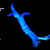
 Invasive breast cancer is detected in left breast as an enhancing irregular mass. The mass is well visualized on reconstructed T1 weighted image. Images and caption courtesy of Soonhoi Ha, PhD, and the ISMRM.
The study findings "pave the way for noncontrast MRI options in clinical and screening settings, potentially broadening MRI use in patients with contrast-related contraindications," Ha and colleagues concluded.
Check out AuntMinnie’s full coverage of ISMRM 2025 here.