4D flow MRI reflects vascular changes associated with the progression of fibrosis in patients with chronic liver disease, according to a study presented at the International Society for Magnetic Resonance in Medicine (ISMRM) meeting.
The preliminary findings are from an ongoing clinical trial in Sweden and suggest that assessing portal vein hemodynamics may identify patients at risk of cirrhosis, noted Shan Cai, a doctoral student at Linköping University, in a May 15 session on functional body imaging.
“We validated 4D flow measurements in the portal vein system against the reference 2D flow MRI, and our results suggest that 4D flow may reflect vascular change associated with fibrosis progression,” Cai said.
Chronic liver disease (CLD), particularly metabolic dysfunction-associated steatotic liver disease (MASLD), is a growing global health concern that can progress to cirrhosis. Portal vein hypertension is common in cirrhosis and can cause high mortality, Cai noted.
While standard 2D flow MRI measures blood flow in the portal vein in a single plane and velocity in one direction, time-resolved, 3D phase-contrast MRI (4D flow MRI) has been shown to be a promising tool, as it provides a more comprehensive and dynamic view. Yet whether the technique can measure hemodynamic changes during fibrosis progression, especially at early stages, remains unclear, Cai said.
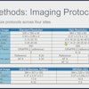
To address the knowledge gap, Cai and colleagues investigated whether 4D flow MRI can distinguish CLD patients at different fibrosis stages from healthy individuals, compared with standard 2D flow MRI. The researchers recruited three cohorts: 37 MASLD patients without advanced fibrosis (18 males), 18 advanced fibrosis patients (17 males), and 18 healthy volunteers. Participants underwent both imaging exams on a 3-tesla scanner (Ingenia, Philips).
Portal vein flow data were analyzed using software (GTFlow, GyroTools), with hemodynamic parameters including average and peak flow and average and peak velocity through one cardiac cycle. In addition, the researchers assessed correlations between flow changes and MR elastography (MRE)-derived liver stiffness.
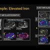
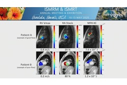
 Velocity streamlines of the portal vein measured using 4D flow MRI in a cirrhosis patient (a 62-year-old male), shown at the cardiac cycle time t = 84 ms. A cut-plane (white marker) matching the 2D flow image plane was used to quantify the 4D flow parameters in the main portal vein.Shan Cai and ISMRM
According to the results, both exams measured significantly increased average flow in MASLD and advanced fibrosis patients compared to healthy controls. The average velocity of the patient groups, derived from 4D flow, was significantly higher than that of healthy controls. In contrast, no significantly increased average velocity of patients was presented in 2D flow MRI.
In addition, among patients with at least significant fibrosis determined by MRE (a cutoff value of 3 kPa7), both 2D and 4D flow MRI showed a significant decrease in average and peak velocity with increasing liver stiffness, according to the findings.
“This confirms an acceptable agreement and strong consistency between 4D and 2D flow MRI in quantifying portal vein hemodynamics in both healthy individuals and patients with chronic liver disease,” Cai noted.
Ultimately, the findings are preliminary, with the study protocol calling for the inclusion of 20 more patients with advanced fibrosis, as well as measurements of portal branches, the superior mesenteric vein, or splenic vein, given the limited spatial resolution of 2D flow MRI in small vessels, Cai said.
“4D flow MRI could serve as an alternative to 2D flow MRI for quantifying portal hemodynamics and may reflect vascular changes associated with fibrosis progression,” Cai concluded.
Check out AuntMinnie’s full coverage of ISMRM 2025 here.