CHICAGO -- Portable, ultralow-field MRI can help clinicians diagnose acute ischemic stroke, according to study results presented December 2 at the RSNA meeting.
The findings could improve patient care by making MR imaging more available, presenter Nandor Kolos Pinter, MD, of the University of Buffalo in New York, told session attendees.
"Neuroimaging is crucial for triage of patients with suspected stroke, and DWI [diffusion-weighted] MRI is highly sensitive to acute ischemic stroke compared with CT," Pinter said. "But access to MRI can be a barrier to its use. Low-field MRI may facilitate detection of acute ischemic stroke in broader populations -- and offer an alternative to how we perform stroke care today."
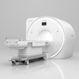
Conventional, high-field MRI ranges from 1.5-tesla to 3-tesla in strength, and is static, bulky, and carries extensive power requirements, Pinter noted. Low-field MRI (64-millitesla) is portable, lightweight, only requires a standard power outlet, and does not require shielding.
The group assessed the performance of low-field MRI for stroke diagnosis via a study called ACuTe Ischemic stroke detection with Portable MR (ACTION PMR) that included 66 patients presenting in the emergency departments of Massachusetts General Brigham, Buffalo General Medical Center, and Ohio State hospitals. The patients underwent diffusion-weighted MR imaging on a low-field MRI scanner (Hyperfine, Swoop 64). Scan time was 17 minutes, Pinter said. Three radiologist readers, each with more than 10 years of experience reading acute stroke MRI, received training on reading the low-field MR images; they then assessed the scans for any lesions and the lesion locations. The researchers evaluated agreement between these radiologist readers by tracking positive and negative predictive values, sensitivity, and specificity.
Pinter and colleagues found the following:
| Predictive values of low-field MR imaging | |
|---|---|
| Measure | Values |
| Sensitivity | 67.5% |
| Specificity | 96.2% |
| Positive predictive value | 96.4% |
| Negative predictive value | 65.8% |
Interrater agreement for DWI lesion detection was kappa 0.80 and for lesion hemisphere was kappa 0.95, Pinter reported.
He acknowledged that low-field MRI's sensitivity and negative predictive value were low but emphasized that the technology's specificity and positive predictive value were high.
"[The low sensitivity could be due to] lesion size, signal homogeneity, and single direction DWI," he said. "But low-field MRI [remains] a very promising tool [for stroke diagnosis and care]."
More research is needed, including tackling how to improve low-field MRI's spatial resolution and developing multidirection DWI techniques, Pinter concluded.
For full 2024 RSNA coverage, visit our RADCast.
