AI is lacking in interventional radiology (IR) and will persist unless more imaging data is saved rather than deleted, according to Benedikt Michael Schaarschmidt, MD, of University Hospital Essen in Germany.
Schaarschmidt's keynote close to a set of IR scientific sessions today pointed out that most AI-related publications focus on pre-interventional imaging because of a lack of data for other use cases, such as in peri-interventional imaging and the interventions themselves.
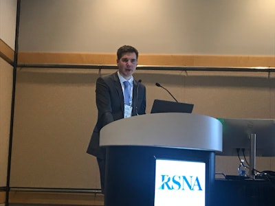 Benedikt Michael Schaarschmidt, MD.Liz Carey
There are two primary reasons why AI is lacking in interventional radiology, according to Schaarschmidt. One reason is the significant shortage of data and integrated datasets. Loads of data generated during interventions are deleted, he said. Annotated data is also sparse.
"The biggest concern is highly variable examination protocols," Schaarschmidt added. "Looking in your same hometown, everybody does their own procedure and when everybody does his own procedure it is really hard to compare data from different sites."
The other obstacle for AI in IR involves problems with data management and a lack of structured reporting, Schaarschmidt explained. Structured data collection is only employed in selected hospitals, he said, and there are still no vendor-independent solutions that collect all the data at the local level.
In addition, useful public datasets like the ones used elsewhere in radiology AI research and development are not available.
"When we talk about national or international databases, we have a really big need for these databases to aggregate interventional data, so the only thing to do is collaborate," Schaarschmidt said.
During his talk, Schaarschmidt cited a retrospective German study published in May that analyzed 754 hepatocellular carcinoma (HCC) patients from six tertiary care centers (between 2010 and 2020). All were to undergo transarterial chemoembolization (TACE).
"In clinical reality, patients with intermediate-stage disease are a heterogenous group with remarkable variations in tumor burden and remaining liver function ... and the patient heterogeneity makes it exceptionally difficult to perform risk scoring and prognosis prediction in patients treated with TACE," the research noted. "To achieve a more holistic picture of patients, and to have an objective indicator of patients’ remaining capacities, body composition assessment (BCA) parameters have become vital as opportunistic imaging biomarkers in patients with hepatobiliary malignancies."
For the study, researchers investigated a fully automated AI-based quantitative 3D volumetry tool to evaluate the roles of different BCA parameters. To collect data, researchers used an infrastructure platform (provided by the German Cancer Consortium) to distribute the software to each site for federated data analysis.
Inclusion criteria included but were not limited to the following:
- Histologically or image-based HCC diagnosis according to the European Association for the Study of the Liver (EASL) criteria
- No treatment performed prior to TACE
- No liver transplantation or tumor resection during the follow-up period after TACE
- Pre-interventional abdominal CT scan availability for BCA parameter calculation
- Full availability of clinical, laboratory, and imaging data
They used automated AI to show that patients with less skeletal muscle volume performed worse than patients who had more muscle volume, and the same was true with total adipose tissue, Schaarschmidt noted.
"You have a really interesting predictor that you can only identify by passing loads of data," he said, adding that the only approach to perform high value interventional radiology is to "think big." Moreover, "projects like this are really great to form broad databases for AI projects in interventional radiology."
During the question-and-answer period, Schaarschmidt suggested videos during interventions be saved and used to assess quality. Ultimately, when it comes to IR more data should be saved, he said.
For full 2024 RSNA coverage, visit our RADCast.


