Monday, November 27 | 11:30 a.m.-11:40 a.m. | SSC14-07 | Room S503AB
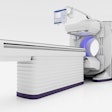
In this talk, researchers from the University of Alabama at Birmingham Hospital will discuss their development of a chart listing technique for optimal radiation exposure parameters for osseous surveys of infants using portable digital radiography (DR).The hospital recently installed a new digital radiography system (Revolution DRX, Carestream Health) in its neonatal intensive care unit. The facility quickly developed optimal technical parameters for chest and abdominal studies for different sizes of babies, to keep radiation dose as low as reasonably achievable.
But staff members at the institution soon began noticing that bone images were missing their targets, with 11 of 29 infant bone images exceeding the institution's target exposure index (EI) range for radiation dose. So, they tasked a group led by presenter Loretta Johnson, PhD, with developing a chart to set parameters for osseous surveys.
The researchers started by reviewing 200 images of infant bone exams to note image quality and the Carestream EI used in the studies. They then performed a series of studies with phantoms selected to simulate infant bones and tested different exposure parameters for image quality. Studies appeared most clear at 40 kVp to 55 kVp, Johnson told AuntMinnie.com.
These tests yielded a range of mAs for each kVp and phantom thickness that produced images within the group's targeted EI range. The researchers then correlated infant bone exams by body-part thickness, kVp, mAs, and exposure index to narrow their desired technique ranges.
At the time of the abstract's publication, 50 infant bone images had been acquired, with only one exceeding the group's targeted exposure index range.
In addition to presenting the group's experiences, Johnson will offer guidance on how institutions can perform their own studies of their radiographic equipment to develop optimal exposure parameters. In future work, she plans on improving image quality for portable chest radiographs of obese adults in the intensive care unit.
