Monday, November 27 | 12:15 p.m.-12:45 p.m. | PD211-SD-MOA2 | Lakeside, PD Community, Station 2
Could digital tomosynthesis using a digital x-ray system with a carbon nanotube x-ray source serve as a better option than conventional x-ray for imaging patients with cystic fibrosis? Find out in this Monday afternoon poster presentation.A team from the University of North Carolina (UNC) at Chapel Hill led by Dr. Elias Gunnell noted that cystic fibrosis is one of the most common life-limiting genetic disorders. Lung disease is a major cause of mortality in these individuals, and repeated infections can damage lung tissue.
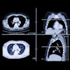
While conventional radiography is commonly used to detect signs of this damage, digital tomosynthesis could offer a better alternative. At UNC, researchers have developed a tomo system that uses a carbon nanotube x-ray source.
Most commercially available digital tomo systems involve an x-ray tube head that pans across a region of interest, acquiring multiple slices of data. But the carbon nanotube system being developed at UNC allows rapid acquisition of multiple images without mechanical motion. The UNC system acquires 31 projections over 10 seconds.
The researchers used the system for cystic fibrosis patients ages 19 to 58 who were scheduled for conventional chest x-ray. Three board-certified pediatric radiologists rated the image quality of the conventional and tomo images and then scored all images for disease severity.
The carbon nanotube tomo system had better scores than conventional x-ray for proximal bronchi, small airways, and vascular patterns, Gunnell and colleagues concluded. They found no difference for lung fields, the trachea, and the diaphragm.
The carbon nanotube tomosynthesis system could be useful for evaluating cystic fibrosis and could enable early detection in pediatric patients, according to the group.


