Thursday, November 30 | 12:45 p.m.-1:15 p.m. | PH263-SD-THB5 | Lakeside, PH Community, Station 5
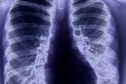
In this poster presentation, researchers from Belgium will describe their efforts to develop more-personalized radiation doses for posteroanterior chest radiography studies using information on body habitus that is available in the DICOM header of images.The amount of radiation dose required to image patients is frequently calculated incorrectly, especially for obese patients, according to presenter An Dedulle of Qaelum, a software developer based in Leuven who is working with the University of Leuven on this project.
Much effort has been put into developing better methods for calculating CT dose, but less attention has been paid to radiography dose, due in some measure to the difficulty in estimating patient size, Dedulle told AuntMinnie.com.
The researchers created a method for estimating patient size for conventional radiography by developing a formula in which dose-area product (DAP) was divided by the exposed area to derive the approximate input air kerma (Aexp) to the patient; the exit air kerma is approximately equivalent to the exposure index (EI). The group, therefore, postulated that the ratio of EI to DAP/Aexp would be related to patient thickness.
To test their hypothesis, the researchers analyzed data from 179 patients who underwent chest radiography and chest CT exams. The gold standard was considered to be the water-equivalent diameter (WED) as calculated from axial CT images.
For the chest radiographs, the researchers extracted DAP, Aexp, and EI from the DICOM header using radiation dose management software (Dose, Qaelum). They calculated the ratio of EI to (DAP/Aexp) against the water-equivalent diameter calculations from the CT scans and applied a curve fit technique to analyze the data. They then validated the methodology on 53 new patients who had chest x-ray and CT exams, calculating water-equivalent diameter derived from the curve fit and comparing it with the true WED derived from the CT data.
Dedulle and colleagues found that their formula of EI to (DAP/Aexp) correlated well with the water-equivalent diameter of the patients from the CT scans, with the best fit occurring with the R2 = 0.89 formula. With the 53 new patients, the mean percentage difference between the group's formula and the CT-derived WED calculations was only 2%.
The results indicate that the technique for calculating patient body habitus could be applied to 2D projection radiographs using simple parameters captured from the DICOM header by radiation dose management software.
"Now, we have for conventional x-ray exams a method to estimate patient size in terms of an attenuation-based metric," Dedulle told AuntMinnie.com. "All data is available to our dose monitoring system and could, therefore, be implemented for automated use in practice."


