Tuesday, November 27 | 3:40 p.m.-3:50 p.m. | SSJ21-05 | Room N229
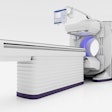
When patients with lung issues in the intensive care unit (ICU) are not stable enough for transport to a CT scanner, a stationary digital chest tomosynthesis system may be a better option than a portable chest x-ray.That conclusion comes from a group of researchers who will describe their experience with a digital chest tomosynthesis system with a carbon nanotube x-ray source array.
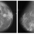
In the study, to be presented by Dr. Elias Taylor Gunnell, currently with Memorial Hospital in Savannah, GA, three thoracic radiologists compared images from a stationary digital chest tomosynthesis system with portable chest x-ray scans. The images came from 22 patients undergoing clinically indicated chest CT and were based on 10 criteria: visualization of lung fields, vasculature, trachea, proximal bronchi, retrocardiac lung, diaphragm, costophrenic angles, ribs, spine, and hardware.
The trio was also asked to assess their confidence in interpreting the scans on a scale of 1 to 7. In addition, they indicated whether stationary digital chest tomosynthesis gave them more information than chest radiographs.
Gunnell and colleagues found that readers had higher confidence scores when using digital chest tomosynthesis for evaluating vasculature, proximal bronchi, and the spine. Two readers gave tomosynthesis higher scores when evaluating the ribs, while one reader offered statistically higher scores for the trachea, retrocardiac lung, and costophrenic angles. Another reader gave chest radiographs significantly higher scores for visualization of the diaphragm.
Confidence scores between the two techniques did not differ significantly for two radiologists, while the third reader gave significantly higher confidence scores to stationary digital chest tomosynthesis. As for additional information from digital chest tomosynthesis, compared with the portable chest x-rays, two readers found additional value 36% and 41% of the time, respectively. The third radiologist said digital chest tomosynthesis added information 95% of the time.
Based on the results, Gunnell and colleagues concluded that digital chest tomosynthesis is a "potentially superior alternative to portable chest x-ray for patients in the ICU setting that cannot undergo CT examination."
