PET/CT reveals a connection between cardiovascular disease (CVD) and cognitive impairment, according to research presented at the recent Society of Nuclear Medicine and Molecular Imaging (SNMMI) meeting.
A team led by Eric Teichner, a medical student at Thomas Jefferson University in Philadelphia found that PET/CT imaging showed "a significant interaction effect on the correlation between F-18 FDG PET and F-18 sodium fluoride (F-18 NaF) uptake in the fusiform gyrus," for example, a region critical for visual categorization.
"Our findings support mounting evidence of the association between CVD and cognitive impairment, demonstrating regional hypometabolism in the presence of increased microcalcification of the carotid arteries," noted presenter Shiv Patil, also a medical student at Thomas Jefferson.
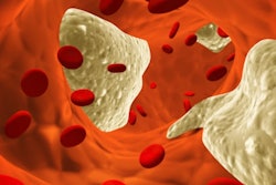
CVD is a leading cause of morbidity and mortality around the world, accounting for 32% of global deaths, Patil noted. The condition is primarily due to atherosclerosis, and it diminishes the delivery of blood and oxygen to organs. When it affects the carotid arteries, it can lead to cognitive impairment and dementia, he explained.
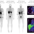
Structural imaging modalities such as CT and MRI can't capture early-stage changes associated with CVD, and that's where molecular imaging comes in: PET imaging can identify vascular and neurological changes before clinical symptoms and structural changes appear, Patil told session attendees. He shared results from a study he and colleagues conducted to assess links between brain metabolism and CVD using PET/CT imaging with F-18 FDG PET to measure cerebral glucose metabolism and F-18 NaF to identify bilateral atherosclerotic calcification, addressing the question of whether there are "differences in brain metabolism in patients with significant molecular calcification."
The research included 104 participants culled from the Cardiovascular Molecular Calcification Assessed by F-18 NaF PET/CT (CAMONA) study; of these, 38 were at risk for CVD and 66 were healthy volunteers. All underwent F-18 FDG PET and F-18 NaF PET/CT imaging. The investigators quantified F-18 FDG PET results with MIMneuro version 7.1.5 (MIM Software) and bilateral carotid F-18 NaF uptake with OsiriX MD software version 13.0.1. They also tracked the relationship between F-18 FDG PET uptake and carotid artery microcalcification.
The group found the following:
- Patient health status showed a significant interaction effect on the correlation between F-18 FDG PET and F-18 NaF uptake in the fusiform gyrus, and this correlation trended directly in patients at risk of CVD but inversely in healthy volunteers.
- Carotid artery microcalcification assessed by F-18 NaF uptake inversely correlated with F-18 FDG PET uptake in the frontal lobe, middle temporal gyrus, and cingulate gyrus.
- F-18 FDG PET uptake directly correlated with average bilateral carotid F-18 NaF uptake in the fusiform gyrus and globus pallidus.
The direct correlations between F-18 FDG PET and F-18 NaF uptake in the fusiform gyrus and globus pallidus may indicate "potential compensatory mechanisms adopted by the brain in response to decreased blood perfusion," Patil noted.
"Future research efforts are needed to validate the clinical application of PET/CT in the detection of arterial calcification and risk of cognitive impairment," he concluded.
