Bronchiolitis obliterans:
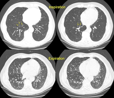
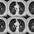
The patient shown below had experienced a severe right lower lobe haemophilus influenza pneumonia the preceding year, but had persistent pulmonary complains. The inspiratory CT images reveal bronchiectatic changes involving the bronchi to the anterior and medial basal segments of the right lower lobe (yellow arrows). Expiratory images deomstrate air trapping in these same segments (white arrows). Air trapping is also evident in the medial segment of the right middle lobe. The findings are consistent with post-infectious bronchiolitis obliterans.