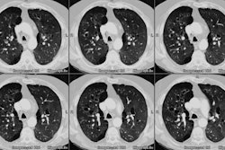
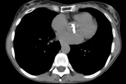
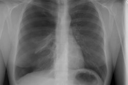
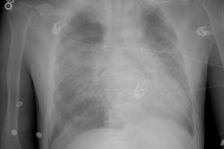
Pneumomediastinum:
View cases of pneumomediastinum
Clinical:
Alveolar rupture, the most common cause of pneumomediastinum,
initially leads to
pulmonary interstitial emphysema. The gas then travels centrally along
the bronchovascular
interstitium to enter the mediastinum. Alveolar rupture occurs
secondary to elevated
intra-alveolar pressure as can be seen in straining (weight lifters,
childbirth), airway
obstruction (mucous plug or foreign body), mechanical ventilation,
coughing, ruptured bleb
with tracking into the mediastinum, crack cocaine sniffing, and blunt
chest trauma (Macklin effect- alveolar rupture with air dissecting
along the bronchovascular sheaths and spreading into the mediastinum
[2]). Tracheobronchial injury and esophageal rupture are less common,
but more serious causes of
pneumomediastinum.
With spontaneous pneumomediastinum, affected patients are usually
asymptomatic, but they may experience chest pain. On physical exam a
peculiar crunching
(or crackling sound- "hamman's crunch") may be heard on auscultation
and varies
with the phase of the cardiac cycle. Palpable crepitation may be felt
over the thoracic
inlet. Although it may produce striking radiographic findings, the
condition is usually
produces little physiologic disturbance. The primary significance of a
pneumomediastinum
is that it may indicated a tracheobronchial or esophageal injury in the
setting of blunt
chest trauma. Pneumomediastinum may progress to pneumothorax if the
mediastinal pleural
ruptures (particularly in positive pressure ventilated patients), but
there is no evidence
to suggest that the opposite (pneumothorax progressing to mediastinum)
occurs.
Tension pneumomediastinum is rare, but occurs when the mediastinal
air produces cardiac compression [3]. The condition can occur in the
setting of mechanical ventilation [3]. The condition can be difficult
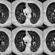
to identify on CXR [3]. ON CT, there will be substantial mediastinal
air and flattening of the anterior cardiac contour, distention of the
IVC, and comrpession of the mediastinal vessels [3].
X-ray:
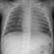
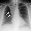
On CXR linear lucencies outline the mediastinal structures and radiate into the neck. The lucencies can extend below the diaphragm to produce a pneumoretroperitoneum. Because of the continuity of the right and left sides of the mediastinum air can outline the central portion of the diaphragm under the cardiac silhouette (referred to as the "continuous diaphragm sign"- a subpulmonic pneumothorax does not cross the midline). On the lateral exam this air will result in visualization of the entire left hemidiaphram (normally obscured by the heart). Other signs indicative of pneumomediastinum include Naclerio's V sign in which gas outlines the lateral margin of the descending aorta and extends laterally between the partietal pleura and the medial left hemidiaphragm (although this sign was originally described in association with esophageal rupture, it is not specific for that condition). In infants, the pneumomediastinum will produce the thymic spinnaker-sail sign.
REFERENCES:
(2) Radiographics 2008; Kaewlai R, et al. Multidetector CT of blunt
thoracic trauma. 28: 1555-1570
(3) Radiographics 2011; Katabathina VS, et al. Nonvascular, nontraumatic mediastinal emergencies in adults: a comprehensive review of imaging findings. 31: 1141-1160
