Whether it’s Alpine, cross-country, or aerial, skiing does a number on the shoulders. Between the pole planting, the skyward maneuvers, and the accidents, mishaps involving the shoulder account for 41% of all upper extremity injuries in these athletes (Sports Medicine, March 1998, Vol.25:3, pp. 201-211).
Specifically, it is the rotator cuff that takes a beating. Rotator cuff strains, and more seriously, tears, make up 20% of shoulder injuries in skiers, according to a study in Clinical Orthopedics (March 1987, Vol.216, pp. 24-28).
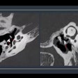
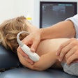
Plain films are good for demonstrating bone abnormalities, such as Hill Sachs or Bankart lesions, while direct visualization of tendons may be achieved by ultrasound or arthroscopic CT. MRI is useful for visualizing the rotator cuff and adjacent bony structures, but can be limited by cost, accessibility, and variable quality (Journal de Radiologie, March 2001, Vol.82:3, Part 2, pp. 317-32).
MRI does offer certain advantages for assessing these types of injuries. In a study at the Mayo Clinic in Rochester, MN, Dr. Eric Williamson and his colleagues used MRI to look at associated rotator cuff pathology with biceps tendon tears.
"One of the primary functions (of) the rotator cuff is the stabilization of the femoral head and the anterior fossa," said Williamson in a talk at the 2001 RSNA meeting. "Tears in the rotator cuff…will frequently cause disruption of the normal orientation of the shoulder joint. This may require surgery. Although the association of subscapularis tears and medial dislocation of the long head of the biceps tendon is well-known, the association of defects of the anterior-posterior rotator cuff with biceps tendon tears is less well-established."
For this study, 126 patients underwent MRI for shoulder pain, followed by arthroscopic or open surgery, between January 1997 and December 2000. Noncontrast imaging was done on a Signa Horizon l.5-tesla unit (GE Medical Systems, Waukesha, WI).
"Standard imaging protocols included coronal and sagittal oblique double-echo spin-echo sequences (typically with a 16-cm field-of-view) with TR = 2000 ms, TE = 20 & 60 ms, NEX = 1, matrix = 256 x 192 and slice thickness/gap = 4/1 mm," Williamson wrote in an e-mail to AuntMinnie.com.
Axial and coronal oblique fast spin-echo sequences were obtained, generally with a 14-cm field-of-view and the following parameters: TR = 3500 ms, TE = 45 ms, NEX = 2, echo train length = 8, matrix = 256 x 256 and slice thickness/gap = 4/0.5 mm, he said.
Finally, axial gradient echo sequences (FOV = 16 cm) with TR of 250 ms, TE of 12 ms, flip angle of 30, NEX of 3, matrix of 256 x 192 and slice thickness/gap of 3/0 mm were employed. Frequency-selective fat-saturated pulses were typically used. The MR interpretations were compared with operative reports.
According to the results, 26 patients were identified as having partial or full-thickness tears of the long head of the biceps tendon. The sensitivity of the MR exam was 60%, while the specificity was 89.9%, and the accuracy 85%.
When a tear was present in the biceps tendon, the prevalence of supraspinatous tears was 96.2%. Infraspinatous tears occurred in 34.6% of the cases and subscapularis tears were present in 50%. No significant relationship was found between the presence, or absence, of a biceps tendon tear and the infraspinatous or teres minor tendon tears.
"Interestingly, the sensitivity of MRI for the detection of biceps tendon tears was somewhat lower than we had anticipated," Williamson said. "Tears of the supraspinatous are more easily detected (on MR), and therefore should heighten the clinical suspicion for a biceps tendon tear."
What MR lacked in sensitivity in the study, it made up for with respectable specificity.
"While it is true that the sensitivity of our MR for the detection of biceps tendon tears was fairly low, our specificity and negative predictive values were fairly high (around 90%)," Williamson told AuntMinnie.com. "One reason for the low sensitivity of MR for biceps tendon pathology may be that the imaging planes chosen reflect the high prevalence of rotator cuff tears. The way we choose to image the shoulder is designed to target the clinically important structures of the rotator cuff that are most likely to be injured. The biceps tendon is not imaged 'in-plane,' and therefore images from multiple sequences must be 'summed' to provide a complete picture of this tendon."
A second study, from the University of Rochester Medical College in New York, came to a similar conclusion regarding the commonality of supraspinatus tears in the shoulder. In this report, 606 cases of rotator cuff tears were mined from a hospital database covering 1995 to 2000. The indications for MRI were shoulder pain, impingement, or ruling out a rotator cuff tear. A combination of tears was seen in 217 patients, and analysis was done on tear location and type, said Dr. Simone Elvey in a presentation at the RSNA meeting.
"Rotator cuff tendon tears generally refer to the tear in the supraspinatus. The question we wanted to answer was: How frequently are other rotator cuff tendons torn? Which subgroups of patients are at risk? What combination of tendon injuries are common, and what are the pre-disposing factors associated with additional tears?" Elvey said.
The researchers found 214 cases of multitendon tears. In this patient population, the most common causes of these tears were rheumatoid arthritis and degenerative diseases of the shoulder, particularly in the glenohumeral joint.
For skiers, the relationship between the glenohumeral joint and the rotator cuff plays a major role in their forward trajectory.
"The rotator cuff is important in normal skiing technique because of the way skiers hold their arms in the skiing stance using ski poles. Rotator cuff injuries occur when the abducted glenohumeral joint is forced into the skier’s side, in spite of strong contraction of the abductors. Impingement can also occur when forces are passed proximally along the humeral shaft as the result of a fall on the elbow," explained Dr. Mike Langaran, a general practitioner with the Aviemore Medical Practice in Aviemore, Scotland. Situated in the Scottish Highlands, the group specializes in treating skiing and snowboarding injuries.
In fact, the newest trend in sports-specific training has skiers concentrating on strengthening their upper bodies, which experts have shown to be the greatest detriment to speed, particularly for cross-country skiers (Newsweek, December 17, 2001).
Langran estimated that shoulder injuries, and rotator cuff strains, account for about 25% of the Alpine ski injuries that he sees at his practice. While the U.K. health system doesn’t automatically call for an imaging exam to diagnoses these injuries, Langran is sure as to the modality of his choice.
"I would have to say that MRI seems to be the best imaging technique if I had to choose," Langran said.
By Shalmali PalAuntMinnie.com staff writer
February 23, 2002
Copyright © 2002 AuntMinnie.com


