CARLSBAD, CA - The simple step of coaching healthcare providers to check CT machine parameters before scanning kids may be one of the best things radiologists practicing in general hospitals can do for pediatric patients, according to Dr. M. Ines Boechat, president of the Society for Pediatric Radiology (SPR) and chief of pediatric imaging at the University of California, Los Angeles.
"The majority of pediatric radiology is practiced by nonspecialists," often in emergency rooms, she said. "It's often only when you look at the parameters that you find out you gave a child an adult dose."
Boechat spoke at the annual meeting of the Society for Pediatric Radiology, which opened in Carlsbad, CA, on Wednesday.
While the risks from CT are still considered small, all agree that exposure should be kept to a minimum.
"The focus is to go to the general hospital and ask: Are you using the adult dose on your pediatric patients? If you over-radiate the patient, your image is no more accurate," Boechat said. "The principle of ALARA -- as low as reasonably achievable -- is what we live by."
Key issues at the six-day conference include how to keep kids from excessive exposure to radiation, the trend toward evaluating groups of results to further evidence-based decision-making, and, for the second year, the emerging use of 3D imaging in pediatrics.
"We have to choose among all the tools do decide which is the best for each situation," Boechat said. "The [Obama] administration is trying to reduce the costs of healthcare and [promote] evidence-based decisions, and going to the literature to analyze results is the best way to do that."
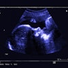
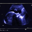
With the enormous increase in pediatric imaging, radiologists have more cases to learn from than ever before. Fetal imaging has grown enormously as well, and many of the studies presented on Wednesday looked at utilizing 3D technology to add value to ultrasound and MR images.
For example, Dr. Archana Malik was able to draw on 438 fetal MRI studies performed at the Children's Hospital of Philadelphia in 2007 to build volumetric 3D images. In a paper presented Wednesday, she concluded that the technology provides valuable pre- and postbirth diagnostic and treatment information.
Malik, a radiologist at Children's Hospital, reported that "3D volumetric-rendered images demonstrate with exquisite detail facial abnormalities [cleft palate] and extremity anomalies [club foot, club hand, arthrogryposis, and neoplasms], providing information difficult to appreciate in two dimensions."
Malik said the ability to turn 2D images into 3D images saves machine time and is of great help in planning treatment, but it has its drawbacks. "It is dependent on the skill of the operator and it takes much time," she said. "It is still a work in progress."
Nonetheless, the information is very valuable, particularly to surgeons. "We try to get 3D imaging on all of our cases," she said. "The 3D images we need involve no motion at all -- that can be tricky."
Dr. Kassa Darge, Ph.D., from the department of pediatric radiology at Children's Hospital, developed 3D images from renal and bladder ultrasound scans to measure volume -- and with simple math, to determine renal parenchymal volume.
"We are going to replace a functional study with a simple application," Darge said. "We have gone from beautiful images to very practical applications."
Darge said the pediatric department at Children's Hospital developed a software program that produces a functional analysis in about five minutes. "One limiting factor was that you could do MR urography but not the functional analysis," he said. "Now our techs have started doing this."
The use of volumetric 3D has proved valuable to surgeons, both in deciding whether to operate and during surgery, he said. And it is safer for the patient. "It is our obligation as pediatric radiologists to utilize this technology to reduce radiation exposure in children," he said. "We should use it to reduce [the use of] other, more invasive diagnostic tools."
One advantage to 3D imaging is that it's based on existing images, according to Dr. George Taylor, radiologist in chief at Children's Hospital Boston. "Some studies show you can reduce the [imaging machine] time," Taylor said. "That's particularly important for pediatric and fetal radiology -- the less you touch a fetus, the better."
When pediatric CT is indicated, Taylor is a fan of 320-detector-row CT, which acquires an entire 16-cm volume of data in one rotation without the need to reconstruct overlapping slices.
"In kids, that means you can use less sedation, you can do functional studies, and you can use a lower dose of radiation," he said. "It acquires a volume so you reduce the noise."
Taylor said that exposure to radiation is worrisome for children, particularly in cancer treatment. "There are oncology protocols where the patient gets repeated CT scans. Some argue that they are already at risk [for the effects of radiation exposure] and that the additional risk is minor," he said. "But if this is your loved one who is affected, a small global risk is no comfort."
Taylor applauds the emerging body of study of imaging in children.
"Most utilized data has been generated for adults, and the indications for kids are very different," he said. "An MRI for sudden-onset back pain in an adult is not indicated, where for a child it is indicated and the likelihood of finding something actionable is 10% to 20%."
By Marty Graham
AuntMinnie.com contributing writer
April 23, 2009
Related Reading
CT often unjustified in the young; German pediatricians lack dose awareness, March 10, 2009
Radiation dose awareness leads to drop in pediatric CT usage, December 12, 2008
Swiss pediatric CT survey leads to national dose standards, October 23, 2008
Study: Radiologists dial back on pediatric CT settings, October 4, 2008
CT experts grapple with rising concerns about radiation dose, May 14, 2008
Copyright © 2009 AuntMinnie.com

