AGRA - Surgeon Commander Dr Pradipta C. Hande, attached to Indian Naval Hospital - INHS Asvini at Mumbai, is a most unlikely candidate for the fighting navy. But her position is one of the few openings for women in a male-dominated navy.
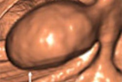
The Indian military is increasingly using the latest imaging technology, and Hande presented a study in which she draws a parallel between the human eyes, which she describes as "windows to the world," and multidetector CT (MDCT) as the imaging specialists' "window to the eyes." She was addressing a session of the 58th annual congress of the Indian Radiological and Imaging Association (IRIA).
"MDCT Imaging of the Orbits" was the presentation topic of Hande's team, which consists of Surgeon Captain Dr J. D'zousa and Surgeon Commanders I.K. Indrajit and R. Pant.
In an exclusive interview with Auntminnie.com, Hande listed three reasons for preferring MDCT:
- Three-dimensional reformatting and volume-rendered imaging is possible.
- Vascular phase studies can be made.
- The process is fast, therefore the modality of choice for an imaging specialist, especially in trauma cases.
It is quite likely the army will find imaging technologies the best method for evaluating orbits. Says Indrajit, "we can now see the path of the bullet, and I am sure my colleagues in troubled areas are using it with effect, probably saving lives in the bargain."
Hande has been using MDCT for evaluating the anatomy of pathologies of the orbits to determine localization and extent of lesions, morphology, intralesion attenuation of characteristics, and paraorbital extension/involvement.
The methods used in cases in which there was a clinical diagnosis on the basis of the signs, symptoms, and ophthalmologic examination were referred for CT scanning wherever indicated when they followed the pattern below:
- Contiguous sections (An advantage of MDCT is high-quality images with thin 1.5- to 2.0-mm sections in spiral protocol.)
- Axial-parallel to infraorbito-meatal line
- Coronal-perpendicular, extending in a posterior direction to include optic chiasm
- IV contrast injection preferred for all studies
- Bone/soft-tissue window for dressing
High-resolution CT imaging of the orbits with MDCT techniques gives excellent delineation, Hande said. "Postcontrast scans enhance the utility of CT scanning, and helps immensely to optimize the diagnosis," she added. This method is presently indispensable as an imaging modality, and it will continue to expand, driven by advancing technology and clinical applications contributing to more specific diagnosis of orbital disease, Hande concluded.
By Ravi Damodaran
AuntMinnieIndia.com contributing writer
January 24, 2005
Copyright © 2005 AuntMinnie.com


