CHICAGO - Researchers from major cancer hospitals said the ability to fuse images obtained through positron emission tomography and computed tomography will benefit patients by giving doctors the ability to improve cancer diagnosis and treatment.
“What we are seeing with PET/CT just knocks your socks off,” said Dr. William Strauss, professor of radiology at the Weill Medical College of Cornell University, New York.
In a series of studies presented during the 88th annual meeting of the Radiological Society of North America, researchers explained their experiences in using the new fusion technology in head and neck cancers, ovarian cancers and lung cancers.
However, Dr. Heiko Schöder, assistant attending physician at Memorial Sloan-Kettering Cancer Center in New York City, said during a press briefing, “ I don‘t think there is any cancer where PET/CT won’t work. I think it has particular advantages in head and neck cancers where there are a lot of small structures that require evaluation.”
In the study Schöder presented, two physicians evaluated images from patients with suspected head and neck malignancies. PET images picked up 154 lesions in 68 patients. Of those lesions 45 were benign, 38 were equivocal and 71 represented malignancies. When PET was combined with CT, using a Siemens-based platform, the doctors judged 62 of the lesions as benign, 18 as equivocal and 76 as malignant. The fusion imaging, Schöder noted, picked up additional areas that were missed by PET alone. “We were able to reduce the equivocal reading by 53 percent, and that was statistically significant (p<0.010),” he said.
Dr. Hans Steinert, assistant director of nuclear medicine at University Hospital Zurich in Switzerland, employing a GE Medical Systems scanner, said that his study of lung cancer patients provided useful information in 40 percent of cases.
He illustrated one case in which the separate PET and CT images were imprecise in determining if a lung cancer had spread. When the fusion image was scrutinized, doctors could quickly determine that the cancer had spread outside the lungs and surgery was no longer a viable option for the patient. Steinert said this case showed the technology could change treatment decisions, and that chemotherapy would be offered to this patient.
“The patients who will benefit the most,” Steinert suggested, “will be those for whom surgery or radiation therapy is planned.” He said the images will be used to guide the surgeon to the exact location of the malignancy and to help the radiation oncologist intensify treatment against the cancer and avoid injury to healthy tissue.
In a third study, Dr. Elliot K. Fishman, professor of radiology and oncology and director of diagnostic imaging and body CT at the Sidney Kimmel Comprehensive Cancer Center at Johns Hopkins Medical Institutions, Baltimore, showed how the fusion images could improve diagnoses of ovarian cancer. “The best imaging study we can do for the detection of ovarian cancer is PET/CT, which has a very high sensitivity for detecting disease and is very specific,” he said.
For the study, performed on a GE platform, 10 PET and 33 PET/CT examinations were performed on 28 patients. The study found that PET alone had a sensitivity of 100 percent and a specificity of 50 percent, while combined PET-CT studies had sensitivity of 73.6 percent and a specificity of 100 percent. The findings indicated that PET/CT appeared to increase the specificity for detecting peritoneal metastases from ovarian cancer over PET alone, Fishman said, and that PET/CT had high overall diagnostic accuracy.
“The excitement of the technology is not just what can be done now but what will occur in the short term,” said Fishman.
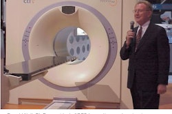
Strauss, who is also clinical director of the nuclear medicine service at Memorial Sloan-Kettering, said all the major scanner manufacturers are developing PET/CT fusion systems. He also said that oncologic imaging is not the only application for the fusion technology, suggesting a major role might be available in cardiology and other areas.
PET/CT fusion combines CT’s structural information with PET’s metabolic information in one set of images, enabling small lesions to be detected with PET and precisely located using CT.
By Edward Susman
AuntMinnie.com contributing
writer
December 3, 2002


