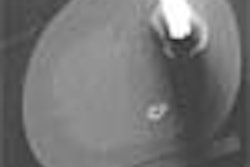
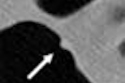
Using a ratio of peak attenuation to kidney stone size on spiral CT allows physicians to distinguish predominantly uric acid calculi from predominantly calcium oxalate calculi, according to a presentation at the 2003 American Urological Association (AUA) meeting in Chicago.
The ability to differentiate uric acid stones from other stone types could be helpful in selecting the appropriate therapy, explained Dr. Patrick Lowry, an endo-urology resident at the University of Wisconsin Medical Center in Madison.
"Uric acid stones can usually be treated with medication and generally don’t require surgery. So if we can predict those stones, we can predict which patients can be managed medically," he said.
The study included 235 patients with flank pain who underwent CT scans on a four-slice spiral unit from GE Medical Systems (Waukesha, WI). The slice thickness was 5 mm. Imaging revealed a urinary tract calculus, and the patients subsequently had passage or extraction of the stone, which was then analyzed for composition.
Each CT scan was analyzed by a single radiologist who determined the size in millimeters, and peak attenuation in Hounsfield units (HU) for each calculus. The ratio of stone size in millimeters to peak attenuation was compared to the stone analysis results to further enhance the accuracy of prediction.
Overall, 145 of the calculi were predominantly calcium oxalate in composition, while 26 were predominantly uric acid. The HU peak unit alone was a significant predictor of uric acid stone type (p=0.0008), Lowry said.
Using a peak attention-to-stone-size ration cutoff of 80, predominantly uric acid stones could be correctly distinguished from predominantly calcium oxalate calculi with 81% sensitivity, 90% specificity, 58% positive predictive value (PPV), and a 96% negative predictive value (NPV).
Using the same cutoff value of 80, and limiting the cohort to those with stones 4 mm and larger, predominantly uric acid stones could be correctly distinguished from predominantly calcium oxalate calculi with a sensitivity rate of 94%, specificity of 90%, a 64% PPV, and a 99% NPV.
Using the same cutoff value of 80 with all stone sizes, predominantly uric acid stones could be correctly distinguished from all other stone types with a sensitivity rate of 80%, specificity of 85%, a PPV of 88%, and an NPV of 98%.
This spiral CT test would be particularly beneficial for patients who had never had a prior kidney stone, Lowry said.
"I am talking about the patient who comes to the emergency room and is found on CT scan to have a stone that is predicted to be a uric acid stone," he said. "If that’s the case, we can give the patient the proper medication in the emergency room that will help dissolve the stone. The patient can then be followed up with medical management to prevent additional uric acid stones."
By Jill Stein
AuntMinnie.com contributing writer
May 19, 2003
Related Reading
Lowering CT dose yields usable results in renal colic, May 24, 2002
One-stop evaluation with novel CT urography approach on modified machine, November 27, 2000
Studies show CT vanquishing intravenous urography in flank pain evaluation, May 1, 2000
Copyright © 2001 AuntMinnie.com


