CHICAGO - Toshiba America Medical Systems lobbed an unexpected shell into the multislice CT wars at this week's RSNA meeting, debuting Aquilion One, a CT system based on 320-slice architecture.
Toshiba had widely been expected to use this week's RSNA show to focus on its 256-slice technology, which the firm has been touting as a work-in-progress for the past several years. But while it was singing the virtues of 256-slice CT, the company was actually planning to make the 320-slice system its next step forward from 64-slice architecture.
 |
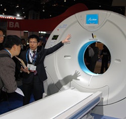
| Toshiba is debuting its AquilionONE 320-slice CT system in its RSNA 2007 booth. |
Toshiba made a number of improvements to its Aquilion architecture to develop the new system. The platform employs the next generation of its Quantum detector technology, Quantum V, with 0.5-mm detector elements, as well as a new tube, called MegaCool V.
Toshiba is calling the unit a dynamic volume CT scanner, due to its ability to capture a large volume of data -- 16 cm -- per gantry rotation. This volume coverage enables the system to visualize an entire organ in a single rotation, without having to stitch together images from multiple passes.
Toshiba believes the dynamic volume approach has multiple advantages. One is the elimination of stitching artifacts, while another is lower radiation dose due to the elimination of overlapping CT slices. The company believes the system produces 80% less dose than a 64-slice model.
Aquilion One's broader coverage area will deliver benefits across a wide range of clinical applications, according to the company. Cardiac and brain perfusion studies will be improved due to the scanner's ability to visualize organ and blood flow dynamically. In brain imaging, for example, Aquilion One can perform an arterial, venous, and whole-brain perfusion exam in a single study and with less contrast and radiation dose.
Toshiba also addressed the data volume issue with Aquilion One, thanks to a specialized workstation developed for the scanner by partner Vital Images of Minnetonka, MN. Scans can be reconstructed in as few as 10 seconds, according to the company, and many noncardiac exams actually have less data than a 64-slice scan due to the lack of overlapping slices.
Aquilion One will begin shipping in the summer of 2008, Toshiba said.
Toshiba also introduced new automated workflow technologies and new clinical applications for the firm’s Aquilion CT product lines. New clinical applications such as Sure CardioProspective gating software that is intended to reduce radiation dose during cardiac exams, and Variable Helical Pitch (vHP) scanning that is designed to permit non-stop helical scanning were unveiled as well.
MRI
The big highlight in the MRI section of Toshiba's booth is Vantage Titan, an enhancement to the company's Vantage family of scanners. Titan features a 71-cm patient aperture and extended 55 x 55 x 50-cm field-of-view, and also has the same 1.4-meter magnet bore length and Pianissimo gradient noise reduction technology as the older Vantage scanners.
Toshiba is touting the power of the scanner's gradients, rated at 30 milliTesla/meter (mT/m) maximum amplitude and 130 mT/m/msec slew rate. Another highlight is the homogeneity of the scanner's magnet, rated at a guaranteed homogeneity of 2 parts per million (ppm) over a 50 x 50 x 50-cm diameter spherical volume (DSV).
Toshiba is making its full range of MR imaging applications available on Titan, including the company's noncontrast imaging techniques such as its recently introduced time and space angiography (TSA) technique. Owners of the company's Excelart Vantage and Excelart Vantage Atlas scanners can upgrade to Titan.
Titan is being shown as a work-in-progress, and has not yet received U.S. Food and Drug Administration (FDA) clearance.
Also being shown as a work-in-progress is a 3-tesla version of the company's Vantage platform. The scanner has gradients rated at 30 mT/m maximum amplitude and 200 mT/m/msec slew rate, and like Titan includes Pianissimo gradient technology.
Toshiba is touting the high homogeneity of the scanner, at 1.6 ppm over a 50 x 50 x 50-cm DSV, as well as its short magnet bore. The scanner will include whole-body imaging protocols, the company said.
Finally, Toshiba is promoting enhancements to the Vantage Atlas system, including its contrast-free MR angiography techniques. The company is highlighting the scanner's 128-element architecture, as well as its different-sized coil elements, which enable improved flexibility in patient positioning.
X-ray
RadRex-i is a new radiography table that features dual digital x-ray detectors, outfitted in a wall stand and table configuration. The system sports a highly automated design, with features such as autotracking, which synchronizes the tube and detector to avoid the need to manually position the system. An autocollimation feature selects the correct collimation for the patient's body part, while an autoprogram feature chooses the correct program for each x-ray procedure, and autopark raises the x-ray tube out of the way after an exam.
RadRex-i also features a patient table with a 600-lb weight limit, a more powerful x-ray tube, and 80-kW x-ray generator. Shipments of the system begin in February.
 |
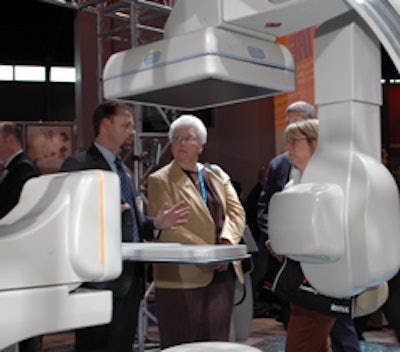
| The Infinix VF-i/BP biplane x-ray system features a multiaxis, floor-mounted C-arm and suspended omega-arm designed for head-to-toe patient access. |
The x-ray system, which is suited for vascular and neuro x-ray procedures, features two flat-panel digital detectors -- a 12 x 16-inch detector on the multiaxis C-arm and an 8 x 8-inch detector on the ceiling on the omega-arm. Infinix VF-i/BP currently is available and shipping.
In addition, a new low-contrast imaging feature, 510(k) pending, will become available for all Toshiba Infinix-i large-panel x-ray systems. Low-contrast imaging is used to provide visualization of soft tissue in the angiography suite.
Ultrasound
Among the highlights in ultrasound is Toshiba's new 4D volume imaging applications for its Aplio and Xario ultrasound systems, as well as new 4D transducers and its upgraded Xario XG system.
Toshiba added Aplio features to Xario to create Xario XG, which has a 19-inch monitor (expanded from the previous 15-inch monitor), as well as standard software features and the new 4D transducers.
Standard software features on Xario XG include:
- Advanced dynamic flow, which is designed to display blood flow with directional information, even in very small vessels
- ApliPure, which uses real-time spatial and frequency compounding technology for enhanced image quality
- QuickScan autooptimization for 2D and Doppler imaging
Toshiba's new line of 4D transducers is designed to help physicians in clinical applications, such as transvaginal/obstetric, small parts, and abdominal imaging. The transducers are currently available.
A microconvex transducer is intended for abdominal applications and R/F ablations, while an endocavity transducer provides volume data for both male and female imaging, including transvaginal/obstetric procedures and prostate imaging. A linear transducer is designed for imaging small body parts, including the testes, thyroid, and breasts.
Finally, Toshiba has teamed with McKesson of Richmond, British Columbia to develop the Ultrasound Multiview Off-line Review Workstation software package, which is used to load 4D data acquired from the Aplio XG and Xario ultrasound systems into a McKesson PACS workstation to enable browsing and reconstruction activities off-line. This feature dedicates the use of the ultrasound system to the acquisition of images, while freeing it from post-processing needs.
By Brian Casey and Wayne Forrest
AuntMinnie.com staff writers
November 26, 2007
Related Reading
Toshiba launches contrast-free MRI technique, November 15, 2007
Toshiba to sponsor new 64-slice CT study in diabetics, November 6, 2007
Road to RSNA, Ultrasound, Toshiba America Medical Systems, November 5, 2007
64-slice MDCT highly accurate for stenosis detection, November 5, 2007
Road to RSNA, MRI, Toshiba America Medical Systems, October 29, 2007
Copyright © 2007 AuntMinnie.com


