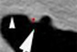
Polyps in the ascending colon can mimic mobile contents such as fecal matter when patients turn from prone to supine position at virtual colonoscopy, potentially skewing interpretation, a new study indicates. Simple awareness of the phenomenon can go a long way toward helping radiologists avoid misinterpretation of virtual colonoscopy results.
The phenomenon partly depends on the degree of abdominal compression, and it can be alleviated by placing a cushion under the lower chest and abdomen during prone virtual colonoscopy (also known as CT colonography or CTC), wrote Ji Yeon Kim, Seong Ho Park, and colleagues from Asan Medical Center in Seoul (European Radiology, February 2011, Vol. 21:3, pp. 353-359).
"Readers of CTC should be aware of the possibility of ascending colon rotation in CTC in order to avoid misinterpreting a true lesion as a pseudolesion," the authors wrote. "Such pitfalls can be avoided by carefully checking the lesion location relative to other fixed points of reference such as the teniae, diverticula, or the ileocaecal valve."
Pedunculated polyps and lesions in mobile colonic segments can be misinterpreted as pseudolesions because they can mimic mobility when the patient changes position, according to the authors. Colonic segments considered mobile include the sigmoid colon, transverse colon, and cecum. However, lesions have been found to move even in the ascending and descending colon segments, which are generally considered immobile, they wrote.
"We have experienced several cases where a fixed lesion in the straight midportion of the ascending colon mimicked mobility due to external rotation of the ascending colon when the patient moved from the supine to the prone position during CTC examination," Kim and colleagues wrote of the problem.
The degree and patterns of ascending colonic rotation have not been well studied; doing so could help avoid misinterpretation of CTC studies, they wrote. The group's colonoscopy database included 4,500 CTC exams conducted between 2006 and 2009, which yielded 37 patients with 43 eligible lesions that fulfilled the study criteria:
- Colonoscopy-proven sessile polyps 6 mm or larger in the straight midascending colon
- Lesion visualization in both supine and prone CTC
- Optimal colonic distension
The 43 polyps included 35 tubular adenomas, three tubulovillous adenomas, two hyperplastic polyps, two inflammatory polyps, and a serrated adenoma.
Virtual colonoscopy exams were acquired prone and supine after one of three cathartic bowel preps, using either sodium phosphate, magnesium citrate, or polyethylene glycol, and automated colonic insufflation with CO2 (ProtoCO2l, Bracco Diagnostics). Images were acquired using a 16-detector-row scanner (Somatom Sensation 16, Siemens Healthcare) with a slice thickness of 1 mm reconstructed at 0.7-mm intervals.
To reduce abdominal compression and avoid poor colonic distention in the prone position, a cushion was placed under the lower chest and pelvis in five obese patients.
The radial location of each polyp was determined by a single experienced reader. A coordinate system was designed to designate the polyp radial location (0° to 360°) along the luminal circumference that was unaffected by rotation of the torso. The degree and direction of polyp radial location change (i.e., ascending colonic rotation) between supine and prone positions correlated with anthropometric measurements, the authors wrote.
The radial locations of the 43 polyps were measured on the supine images, then a month later on the prone images. The difference between the two positions was registered as a value between -180° (internal rotation) and 180° (external rotation) of the colon with the patient in the prone position compared to supine. The same measurement process was repeated a month later to assess reader variability between the two measurements.
"The ascending colon was usually found to rotate externally as patients moved from supine to prone positions, partly dependent on the degree of abdominal compression," Kim and colleagues wrote.
The results showed changes in the radial polyp location ranging from -23° to 79° (median, 21°) due to movement between prone and supine positions, showing external rotation of the ascending colon in almost all cases. Ascending colon movement ranged from 2° to 79° in 36 of 37 patients and in 42 of 43 lesions. The degree and direction of rotation correlated modestly (degree of rotation: r = 0.427, p = 0.004; direction of rotation: r = 0.404 p = 0.007) with the degree of abdominal compression in the anterior-posterior direction in prone position.
"Although these degrees of ascending colon rotation would not mimic a complete movement with gravity of a pseudolesion from the dorsal to the ventral wall (i.e., approximately 180° change), such rotation can still mimic sticky stool pieces which are partially mobile, and may result in diagnostic errors," Kim and colleagues wrote.
For lesions on the dorsomedial side of the ascending colonic wall in particular, external rotation will make them appear on the ventromedial side, they wrote, making them appear to have migrated in the direction of gravity and raising the false impression of a mobile stool.
A large degree of ascending colon rotation was seen in some patients, possibly due to large differences in the length of the mesocolon that are not completely embedded in the retroperitoneum. Body mass index and abdominal circumference were not correlated with either the degree or direction of ascending colon rotation, although large body habitus is generally associated with poor colonic distention in the prone position caused by abdominal compression, the authors noted.
As for study limitations, the possibility exists that some pseudolesions may have been misinterpreted. Also, the movement of the descending colon was anticipated to be similar to that of the ascending colon, but the absolute movement was not confirmed as comparing the lesion location with the locations of three colonic teniae is difficult, they wrote.
"The present findings indicate that readers of CTC should be aware of the possibility of ascending colon rotation in CTC in order to avoid misinterpreting a true lesion as a pseudolesion," the group concluded. "Such a potential CTC pitfall can be avoided by carefully checking the lesion location relative to other fixed points of reference such as the teniae, diverticula, or the ileocaecal valve. The risk of misinterpretation may also be reduced by using measures that decrease or prevent abdominal compression in the prone position during CT, such as placement of a pillow or a cushion under the lower chest or the pelvis of the patient."


