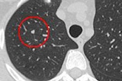
For surveillance of previously detected lung nodules, CT radiation dose can be cut to approximately 3% of the original exam dose, concludes a new study in the September edition of the American Journal of Roentgenology.
The dose savings are important because lung cancer screening patients are typically scanned several times over the course of CT surveillance. Still, missing a growing lesion is serious business, so the technique will need additional research data before the lowest doses can be implemented clinically.
Researchers from Stanford University and the University of Bern in Switzerland found no significant difference between the number of lesions that could be visualized at 10 mAs versus the standard tube-current setting of 300 mAs (AJR, September 2011, Vol. 197:3). The ultralow-dose protocol translates into a substantial reduction in radiation risk, according to the study's lead author, Dr. Andreas Christe.
"The risk of radiation-induced cancer death for one standard chest CT is estimated at roughly one in 4,000 and could be reduced hypothetically to one in 120,000 with this low-dose protocol," he said in a statement accompanying release of the study.
The study involved three radiologists who each examined 50 patients who had been scanned at different dose levels. When reviewing scans of patients imaged with the 10-mAs protocol, the readers did not show a significant difference in sensitivity versus when they reviewed images of patients scanned at the standard 300-mAs setting, Christe said.
The radiologists were able to identify 88% of the lung nodules at 10 mAs, compared with an average of 91% at 300 mAs, the group found. Specificity at the standard 300 mAs setting was 92% and did not decrease when the dose was reduced.
Pending the results of a developing clinical study, follow-up patients at Christe's institution will be scanned at 40 mAs. The present study has limited applicability because it used a smaller field-of-view than normal in clinical practice, as well as dose simulation, the authors said. For now, a setting of 40 mAs should be used going forward because it decreased the measurement variability compared to radiologists performing the measurements manually.
"It is essential that we can get exact measurements of the nodules so we can determine if the nodule has grown between CT exams," Christe said.


