CHICAGO - If cost isn't an issue -- and that's a big if -- it's time to say goodbye to chest radiography, according to research from Norway presented on Thursday at the 2012 RSNA meeting.
In a study that compared conventional chest radiography to 320-detector-row CT using advanced iterative reconstruction, the team found no advantages to chest radiography outside the realm of cost -- whereas they found enormous advantages in diagnostic power with regard to CT.
In fact, the switch to CT would have been completed long ago were it not for radiography's big advantage: a far lower dose profile. Chest CT has traditionally delivered as much as 4 mSv in radiation from a single scan, compared with about 0.05 mSv for a chest x-ray, said Dr. Trond Aaløkken from Rikshospitalet in Oslo.
"First, CT is more expensive than chest radiography; second, the time spent in the lab is longer for CT than for chest x-ray; and last, the most important point is the dose, which is many times higher for CT than for chest x-ray," Aaløkken said in his presentation. "The goal, of course, is to combine the low-dose of chest x-ray with the image quality of CT, but this is ambitious and has not been possible."
But using 320-detector-row ultralow-dose CT with an advanced iterative reconstruction algorithm might make it possible, he said.
The study aimed to compare image quality, radiation dose, and laboratory time between chest radiography and ultralow-dose chest CT reconstructed with advanced adaptive iterative dose reduction (AIDR 3D, Toshiba Medical Systems).
Aaløkken and colleagues examined 13 patients with suspected pulmonary lesions, following chest radiography with ultralow-dose CT acquired on a 320-detector-row scanner (Aquilion One, Toshiba). Images were acquired at 135 kv and 10 mA, with 0.5-mm slice thickness and rotation time of 350 msec.
Three experienced chest radiologists rated both sets of images for pathological findings on a three-point scale, and image quality was rated according to nine criteria of the European guidelines for chest CT and chest radiographs.
Upon analyzing the results, Aaløkken and colleagues determined the following:
- The average laboratory time was five minutes for chest radiography versus seven minutes for CT.
- The average effective dose was 0.05 mSv for chest radiography and 0.105 mSv for CT.
- The 33 relevant findings included 19 masses, nodules, and micronodules.
- Detection sensitivity for all observers and all findings was 18% ± 3% for radiography and 89% ± 2% for ultralow-dose CT.
- The positive predictive value for radiography was 37%, including 31 false-positive findings, while the positive predictive value for CT was 98% with two false-positive findings.
- Image quality for all readers was significantly higher for the CT images (p < 0.05).
"Both image quality and the ability to detect and exclude pathology was strikingly better for ultralow-dose CT than for radiography," although laboratory time was slightly longer for CT, and CT dose was higher but still minimal, Aaløkken said.
 |
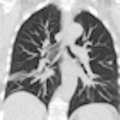
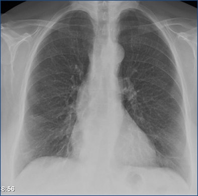
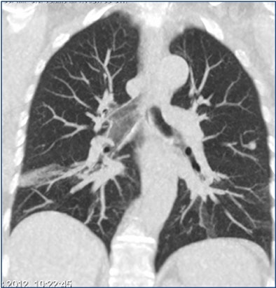
| Above is a false-negative chest x-ray. Below, an ultralow-dose 3-mm axial chest CT of the same patient acquired on a 320-detector-row scanner (Aquilion, Toshiba) and reconstructed with AIDR 3D shows a 7-mm nodule. At bottom, the coronal maximum-intensity projection shows a 7-mm nodule in the left lung and some atelectasis in the right lung. All images courtesy of Dr. Trond Aaløkken. |
 |
 |
The study demonstrated "superior image quality for CT versus radiography," he said. "The similar radiation dose and laboratory time leaves cost as the only reasonable argument in favor of radiography." Otherwise, "it means we can say goodbye to radiography."
Responding to a question from the audience, Aaløkken said that potentially longer interpretation time for CT was not examined but is a valid issue that needs to be addressed.
Session moderator Dr. Narinder Paul from the University of Toronto also noted the possibility of more findings with CT, potentially leading to additional tests, costs, and risks.


