Tests of a prototype photon-counting detector breast CT scanner show that the system could be useful as an adjunct to low-dose breast CT by separating the energy levels of different types of materials within the breast to assess tissue composition and improve cancer diagnosis.
The study team from Duke University Medical Center built the system from off-the-shelf parts including charged integrated flat-panel photon-counting detectors and an x-ray source, and tested it using phantoms containing plastic and biological materials. They presented their work at ECR 2017.
After imaging the phantoms and a cadaver breast, the results showed that the scanner measured energy spectra within established guidelines, and objects placed in the phantom were easily seen in the high-contrast environment. What's more, the system acquired images at a radiation dose comparable to that of film-screen mammography.
"We have reliably imaged geometric phantoms including some biological material," as well as human breasts from a cadaver, said Martin Tornai, PhD, in his ECR talk. Tornai is an associate professor of radiology and biomedical engineering at Duke, and also director of the university's multimodality imaging lab.
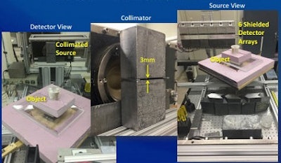 Tabletop layout of photon-counting detector breast CT scanner. All images courtesy of Martin Tornai, PhD.
For all the strengths of mammography and, more recently, digital breast tomosynthesis, both modalities are burdened with the unpleasant requirement of breast compression, a process that has never been required for breast CT, even in prototypes dating back to 1975.
Adding photon-counting detectors to breast CT brings the promise of low-dose acquisition without compression. It also creates the possibility of spectroscopic acquisitions, in which images are acquired based on the different chemical properties of tissue -- which can be useful for differentiating malignant from benign tissue.
A new prototype scanner from Duke could readily be incorporated into the institution's existing dedicated breast SPECT/CT imaging system, according to Tornai.
"We thought it would be nice to use this fantastic technology to acquire mammo images in uncompressed mode," he said in his talk.
The prototype scanner has six 128-element x-ray channels in a 60-cm linear array with cadmium zinc telluride (CdZnTe) digital detectors. This results in an 800-micron pixel pitch and an energy range of 20 keV to 170 keV.
Despite the tube's high energy capacity, the system operates mostly in the range of 30 to 40 keV, Tornai explained.
"Around 40 keV is the energy range we're most interested in because it's tuned to having a high signal-to-noise ratio squared over dose -- [offering] high dose efficiency when we're acquiring CT data with the flat-panel detectors," he said. "With the lower keV, you get quite a nice quasi-monochromatic energy spectrum."
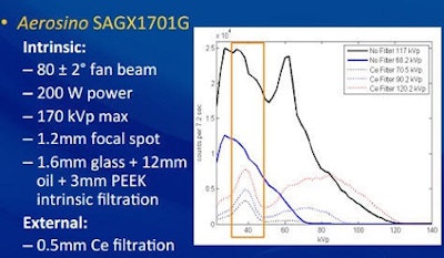 With the Aerosino x-ray source, tube energies of 30 to 40 keV were the most dose-efficient.
To test the system, the Duke group scanned phantoms imaged for 720 projections on the rotation stage at the center of rotation. Phantoms consisted of low and high scatter rods and tubes (1.1 to 4.7 mm in diameter), six different types of materials (acrylic, breast glandular- and fat-equivalent plastics, polyethylene, and oil cylinders), and finally a cadaveric human breast supported in a 12-cm-diameter breast cup, Tornai said.
The results showed that measured energy spectra from 70 to 120 kVp with and without filtration were representative of those in the literature. For both low- and high-scatter phantoms, rods greater than 1.5 mm were easily seen. For the cylinders, measured attenuation coefficients were within 6% of true narrow-beam values and were easily separable in histograms of reconstructions. Reconstructions of breast slices yield glandular, adipose tissues along with skin boundaries.
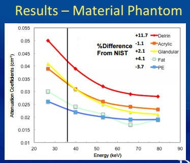 Phantom results for various materials were similar to those of the National Institute of Standards and Technology (NIST).
The energy spectra measurements "show that we can, in fact, measure the attenuation coefficients of individual materials across a variety of energy spectra," Tornai said. "We're within 10% [of published values] except for Delrin, but we don't expect much in the way of high-attenuation materials except for calcifications."
In images of the cadaver phantom, an acrylic bead was easily seen against the glandular background.
Still, the cadaver image wasn't perfect. Some central artifacts and nonuniformity artifacts very common to CT haven't been corrected yet.
"And we're still dealing with circular artifacts -- something we're trying to optimize in data collection," Tornai said.
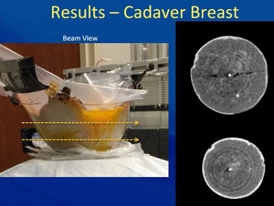 Beam view of cadaver breast shows image artifacts.
As for the results of filtration, the cerium image alone was "quite terrible," he said, adding that it may be necessary to start with the broadband spectrum and then select for certain energies to image.
The study shows that the system reliably imaged geometric phantoms that included biological material, and reliably reproduced attenuation coefficients and properties of materials, Tornai concluded, "although we sincerely think that smaller detectors might be needed, on the order of 100 microns or so for breast CT, and we certainly need a smaller focal spot than the one we have."
In response to a question from the audience, Tornai said that microcalcifications weren't visible because data were reconstructed at 1-mm pixels; however, they could be perceived in a way because the high contrast revealed calcified pixels against the lower-attenuating glandular background.
Once optimized, linear arrays of photon-counting detectors could be used to distinguish tissue composition in low-dose dedicated breast CT, according to the researchers.


