SAN FRANCISCO - Exquisitely detailed ultrahigh-resolution (UHR) CT is on its way thanks to several technologies, although most haven't reached the market yet, according to a talk on Monday at the International Society for Computed Tomography (ISCT) meeting. The technologies are revealing new levels of anatomic detail in several applications. But there are a few downsides, including the potential for higher radiation dose.
In his talk, Dr. Mathias Prokop, PhD, from Radboud University Nijmegen Medical Center in the Netherlands surveyed the technologies that are currently being deployed, from commercial conebeam CT technologies to prototype UHR detector scanners with smaller detector elements and investigative photon-counting CT scanners.
Prokop, a professor of radiology at Radboud University, also looked at a number of clinical applications that seem like a good fit for UHR CT, from high-resolution lung and inner ear images to CT angiography of the coronaries and brain.
"Ultrahigh-resolution imaging is something we have dreamed of for a long time, and with the advent of conebeam CT, our clinical colleagues have seen ultrahigh-resolution arrive, especially in dental imaging and the extremities," Prokop said. "But the question is, can we do that with standard CT?"
An unfinished business
 Dr. Mathias Prokop, PhD, from Radboud University.
Along with the strengths of UHR scanning come a few drawbacks, he said. Take conebeam CT, which brings higher resolution to some applications, but suffers from fewer projections than standard CT and comes with lower detector efficiency, leading to high levels of electronic noise. Conventional CT scanners should be able to do a better job with higher resolution and fewer artifacts than conebeam.
CT resolution depends on the number of unique rays the scanner is sending through the patient, Prokop explained. The number of unique rays depends on the number of detector elements and their thickness.
"In other words, the thinner these rays are the higher the resolution -- and that is determined by the size of the detector elements," he said.
However, measuring spatial resolution is a tricky job that vendors do differently, he said. The modulation transfer function (MTF) is the standard measurement tool. MTF can be reduced to a single curve by applying a cutoff value to the MTF number, be it 20%, 4%, or 0%. But different cutoffs produce vastly different MTF results, Prokop said.
Using about a 20% cutoff gives a good idea of the resolution achieved on the curve, but vendors generally use 4% or 0% in their brochures, which provides little useful information. Going from a cutoff of 20% to 4% brings the line pairs that can be visualized from 21 to 28. Going from 20% to 0% brings the resolution up to 58 line pairs per mm. With measurement differences so large and potentially misleading, the buyer should beware as to how the vendor is measuring spatial resolution.
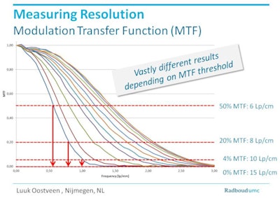 MTF is used to measure spatial resolution, and it varies widely depending on how it is calculated. Image courtesy of Luuk Oostveen and Dr. Mathias Prokop.
Ways to ultrahigh-resolution CT
To get more projections that produce UHR images, you need to use faster detector materials that can read out their information faster, Prokop explained. Alternatively, one can set the gantry to rotate more slowly so the readout is not affected. Another technique is to reduce the size of the detector material by using lead grids that shield half of the detector element. It's not very dose-efficient, but it makes the beam smaller for higher resolution.
Other high-resolution techniques include using smaller detector crystals, or photon-counting detectors, which provide a more narrow response function while delivering pixelation on the side of the anodes rather than on the side of the detector materials, Prokop said.
Conebeam CT
Conebeam CT is used frequently by dentists, and a few other applications such as inner ear imaging have shown excellent results, including resolution of 150 micrometers by a group in Belgium.
"But you see artifacts if you look at larger areas, and the resolution is not the same for all scanners," Prokop said.
In conventional CT, there are ways to "push" the resolution higher, including the use of model-based iterative reconstruction, he said. However, at this point the resolution becomes dose-dependent, so the dose can rise along with the resolution.
UHR detectors
Next in the pipeline are ultrahigh-resolution detectors, built with superfine detector grids for an effective detector element size as small as 0.25 mm x 0.25 mm, along with very thin septae to reduce dead space, Prokop said. Toshiba Medical Systems created such a prototype and is now planning to commercialize a scanner with 0.25-mm detector size.
New detector materials provide for more rapid readout of data. In fact, everything in the imaging chain is optimized to accompany the new detectors, including prefiltering for the x-ray tube, iterative reconstruction, numeric scatter suppression, and binning of detectors for normal resolution mode, Prokop said.
"Basically, everything has to be optimized to get rid of some of the disadvantages of high-resolution imaging, and the main disadvantage is image noise," he said.
Radiologists can see ultrahigh resolution in several anatomic targets with the prototype scanners, including the inner ear, and lung images for assessment of emphysema in ultrahigh resolution at a dose of about 4.5 mSv, Prokop said. Other promising applications include CT angiography of the coronary arteries, brain, mesentery, and run-off vessels, and for presurgical staging before major tumor surgery.
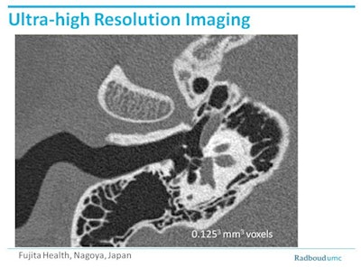 Example from a prototype Toshiba scanner (Aquilion Precision) shows the status of an inner ear at 0.25. Tiny electrodes that could not have been visualized on standard CT are clearly visible. Image courtesy of Dr. Mathias Prokop and Fujita Health, Nagoya, Japan.
"In order to get that, you are ending up with radiation doses that are not excessive, but they are on the higher end of what we're usually using," he said. The UHR scanners are also great candidates for image processing and quantification purposes.
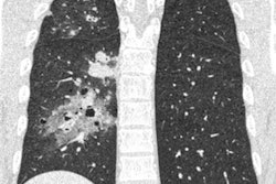
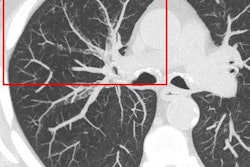
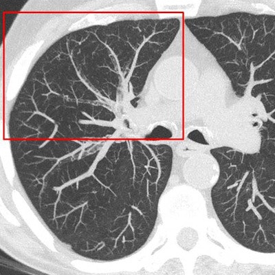
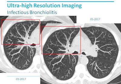 Ultrahigh-resolution lung imaging depicting infectious bronchiolitis on a Toshiba Aquilion Precision scanner shows additional morphologic detail compared with conventional CT. Image courtesy of Dr. Mathias Prokop.
Photon-counting scanners
Photon-counting detector scanners, once commercialized, will provide resolution that is superior to even UHR CT, due to their energy selective organization of CT data in separate bins, Prokop said. Siemens Healthineers is testing its prototype scanners in several locations, including the Mayo Clinic in Rochester, MN, and the U.S. National Institutes of Health in Bethesda, MD.
In photon-counting CT, the photons are converted directly to image data and separated by their energy values. A downside is that energy saturation can be a problem at higher detector doses, he said. As for spatial resolution, photon-counting scanners have slightly worse MTF than normal detectors but far higher signal-to-noise ratio, providing overall better image quality than a conventional system.
In summary, spatial resolution in UHR scanners varies depending on how it is measured, Prokop said.
"You have to be really careful when you compare various vendors," he said. Determine how they measure their spatial resolution and don't trust the numbers until you know: A 20% cutoff is reasonable for MTF, 4% is barely acceptable, and 0% is practically unusable and therefore not acceptable, he added.
Finally, high-resolution works best in anatomic areas with high intrinsic contrast, such as the lungs and bones and in CT angiography applications, and is less favorable in high-absorption regions, Prokop said.


