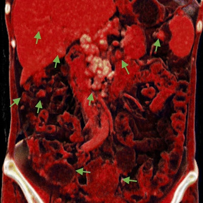
The photorealistic detail provided by cinematically rendered CT scans helped researchers from China characterize and stage ovarian tumors better than they could with traditional volume rendering and multiplanar reconstruction, according to an article published online in the Journal of Ovarian Research.
In the case report, first author Dr. Yi-ren Jin and colleagues from Yunnan Cancer Hospital used computer software (syngo.via, Siemens Healthineers) to convert CT scans of patients with ovarian cancer into cinematically rendered 3D images (J Ovarian Res, September 26, 2018).
Though CT has long served as the primary imaging modality for characterizing and staging ovarian tumors, it tends to provide an inadequate view of tumors that are either poorly vascularized or attached to nearby structures such as the intestines, the authors noted.
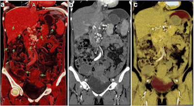
 Cinematically rendered CT scan of the abdomen and pelvic cavity of a patient with ovarian cancer and metastases (a). Multiplanar reconstruction of the same CT scan shows an overall view of the lesions comparable to that of cinematic rendering but with less detail (b). The volume-rendered CT scan does not display the lesions clearly (c). Image courtesy of Jin et al. CC BY 4.0.
Believing that cinematic rendering could help improve the preoperative evaluation of ovarian cancer, Jin and colleagues applied the technology to two clinical scenarios. The first case involved a 44-year-old woman who had previously received treatment for ovarian cancer. Follow-up CT scans two years later revealed that several new nodules and masses had grown around the abdomen and pelvis.
The patient's physicians required a more detailed look at the nodules and masses than conventional CT offered, before being able to determine her eligibility for surgical treatment. To that end, the group used various techniques to visualize the metastases.
Traditional volume-rendered CT scans did not effectively distinguish the metastases from the surrounding gastrointestinal tract. Multiplanar reconstruction was able to display an overall view of the metastases, but only with minimal detail. In contrast, the cinematically rendered CT scans showed that the metastatic nodules had invaded and were deeply adhered to the colon wall. This finding led to the clinicians' recommendation against surgery in favor of chemotherapy.
In a separate case, the ultrasound exam of a 51-year-old woman with abdominal discomfort revealed a pelvic mass. Follow-up CT scans further identified multiple masses near the ovaries. Unlike with traditional volume rendering and multiplanar reconstruction, cinematic rendering of these CT scans clearly depicted the masses and showed that they could safely be removed, even though they were attached to the bladder.
For both patients, cinematic rendering delivered more detailed characterization of the ovarian lesions than did the other methods of visualization. The technique ultimately helped the clinicians differentiate the malignant lesions from benign ones and determine the viability of their surgical removal.
Cinematic rendering may be able to compensate for two main issues with traditional 3D rendering: First, cinematic rendering can distinguish between structures with a much smaller difference in density (within 10 HU) than volume rendering and multiplanar reconstruction can, according to the authors. Second, cinematic rendering allows clinicians to adjust parameters to capture lesions with low vascularity that would otherwise be difficult to see.
"Cinematic rendering with highly detailed and lifelike images is very helpful in preoperational evaluation and surgery planning. ... [It] can provide a more global impression of the spread of ovarian cancer and allow for high-resolution delineation of the structural changes of adjacent and overlying structures," they wrote.


