
Tracking changes in lung holes over time with chest CT in patients with emphysema reveals disease progression and may predict poor outcomes, suggests a study published July 15 in Radiology.
The finding is from an exploratory analysis of CT scans from 108 patients over six years and provides a potential new way to visualize and quantify the dynamic natural course of the disease, noted lead author Yura Ahn, MD, of the University of Ulsan in Seoul, South Korea, and colleagues.
“Emphysema holes change longitudinally in various ways, but current CT measurements lack the ability to fully capture these changes beyond measuring the extent of emphysema,” the group wrote.
Traditionally, emphysema is assessed using pulmonary function testing, specifically forced expiratory volume in 1 second (FEV1). However, this measurement lacks the sensitivity to evaluate short-term changes, the authors explained.
Conversely, quantifying emphysema by measuring the density of lung tissue via chest CT lung densitometry has been extensively validated in relation to physiologic measures, they noted. Still, emphysema holes – empty spaces in the lungs that develop when alveoli are destroyed – vary in diameter and shape over time, and densitometry alone cannot assess these individual dynamics in patients, they added.
Thus, in this study, the researchers aimed to determine whether chest CT can track emphysema holes longitudinally. They sought to group them according to their dynamics and investigate their relationship with change in FEV1, disease progression, and mortality.
The group analyzed data from 108 participants in the Korean Obstructive Lung Disease cohort study from June 2005 to October 2013 who completed baseline and six-year follow-up CT with identical protocols. Significantly, they used deep learning–based software to identify and track the holes based on changes in diameter in 2-mm increments.
According to the results, of the 108 participants (mean age, 63.4 years old; 104 male), 39 had emphysema progression. Enlarged preexisting holes were marginally associated with a greater decline in FEV1 (p = .049), the group reported.
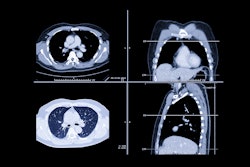
 Emphysema progression with hole changes, with holes colored according to their diameter in 2-mm increments. (A) Baseline inspiratory nonenhanced axial CT scans in a 48-year-old man with chronic obstructive pulmonary disease show emphysema holes with hyperexpansion of secondary pulmonary lobules (white arrows) and thin alveolar walls visible in the right upper lobe. This emphysematous lesion is filled with holes of varying sizes. A single large air cyst was observed in the left upper lobe (black arrows), classified as a single emphysema hole of 20 mm or greater (bright green). (B) At six-year follow-up inspiratory nonenhanced axial CT, the holes in the right upper lobe have merged (white arrows), with no visible alveolar septa remaining, resulting in the formation of a large new hole of 20 mm or greater (bright green). Meanwhile, the hole in the left upper lobe also enlarged, merging with adjacent holes (black arrows). The size of other holes in the paramedian left upper lobe decreased.RSNA
Lastly, participants with 5% or greater volume of increased-diameter holes had worse overall survival (log-rank P < .001), according to the findings.
“Emphysema hole–tracking results showed that a greater volume of holes that increased in diameter were related to change in FEV1, disease progression, and mortality,” the group wrote.
Ultimately, while the study offers a potential way to visualize and quantify the dynamic natural course of emphysema holes beyond changes in densitometry values, external validation with a larger cohort is warranted to confirm the findings, the researchers concluded.
In an accompanying editorial, Edwin van Beek, MD, chair of clinical radiology at the University of Edinburgh, noted that the study moves precision medicine for COPD and emphysema a step forward, particularly given the researchers’ use of deep learning–based assessment.
The development of such technologies provides increasingly sophisticated insights into the heterogeneous group of patients with COPD, which will hopefully facilitate more precise identification of disease phenotypes, he wrote.
“This should enhance the more targeted approach of clinical trials to develop new treatments, which would fulfill an urgent clinical need. Radiology is clearly at the very heart of these developments,” Beek concluded.
The full study is available here.


![Axial images from unenhanced calcium score cardiac CT (left) and curved planar reformation images from CT angiography (right) show that higher long-term exposure to air pollution is associated with greater coronary artery calcium and more obstructive coronary artery disease (CAD). Top row: Images in a 68-year-old male patient with higher 10-year mean ambient air pollution exposure (7.9 μg/m3 for particulate matter measuring ≤2.5 μm in diameter [PM2.5] and 17.4 parts per billion [ppb] for NO2) with extensive CAD (coronary artery calcium score [CACS] >1,000 and obstructive CAD [≥70% diameter stenosis]). Bottom row: Images in a 57-year-old female patient with lower 10-year mean ambient air pollution exposure (6.3 μg/m3 for PM2.5 and 4.6 ppb for NO2) with no CAD (CACS = 0 and no obstructive stenosis).](https://img.auntminnie.com/mindful/smg/workspaces/default/uploads/2026/06/hanneman.r6SMLzkezo.png?auto=format%2Ccompress&dpr=2&fit=crop&h=167&q=70&w=250)


