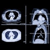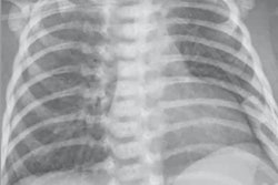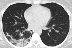
CT imaging data taken from passengers and crew onboard the Diamond Princess cruise ship in Japan showed abnormal lung findings in those with COVID-19, even in asymptomatic patients, according to a study published March 17 in Radiology: Cardiothoracic Imaging.
"In this study, we investigated the chest CT findings in laboratory-confirmed COVID-19 cases in an environmentally homogenous cohort of cruise ship passengers and crew members, comparing the CT characteristics of asymptomatic and symptomatic cases," wrote a team led by Dr. Shohei Inui of Japan Self-Defense Forces Central Hospital in Tokyo. "Noticeably, we found lung parenchymal changes on CT in up to 54% of the asymptomatic cases."
In February, the Japanese government quarantined the American-owned cruise ship the Diamond Princess off of Yokohama, Japan, for two weeks, after a case of COVID-19 was found on board. The ship was carrying about 3,700 passengers. During the quarantine, more than 700 were infected.
"Passengers and crew members of the Diamond Princess cruise ship underwent RT-PCR [nucleic acid testing with reverse transcription polymerase chain reaction] during the quarantine period, and those who showed positive results were transferred to hospitals in Japan," the group wrote. "Among all RT-PCR positive cases from the cruise ship, asymptomatic, mildly symptomatic, or familial clusters with infection among relatives were admitted to the Japan Self-Defense Forces Central Hospital (Tokyo, Japan) for further investigation."
The study included 112 cases of COVID-19 confirmed with RT-PCR. The mean age of the patients was 62. The researchers reviewed the CT images and calculated the severity score for each lung lobe and for the entire lung, then compared the findings between asymptomatic (82 patients, or 73%) and symptomatic cases (30 patients, or 27%).
Of the asymptomatic cases, 54% showed signs of pneumonia on CT, while of the symptomatic cases, 80% had abnormal CT findings. Lung opacities and airway abnormalities were more frequently identified on CT in symptomatic cases than asymptomatic ones, while asymptomatic cases showed more ground-glass opacity predominance over consolidation, the authors noted.
| Lung abnormalities on CT in coronavirus cases | |||
| Characteristic | All cases (112) | Asymptomatic cases (82) | Symptomatic cases (30) |
| Lung opacities | 61% | 54% | 80% |
| Airway abnormalities | 27% | 18% | 50% |
| Underlying lung disease | |||
| Emphysema | 6% | 5% | 10% |
| Pulmonary fibrosis | 3% | 2% | 3% |
CT severity scores were higher in symptomatic cases compared with asymptomatic ones, particularly in the lower lung lobes (both right and left lower lobes, 2 versus 1), according to the authors.
More research is needed, especially because COVID-19 shows abnormal results on CT even in individuals who aren't manifesting symptoms, Inui and colleagues noted.
"Further studies still are warranted to uncover the underlying mechanism responsible for the clinical-radiological dissociation seen in some of the asymptomatic COVID-19 cases, as well as to determine the impact of this finding on clinical decision-making," the group concluded.


