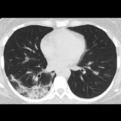
A study conducted by researchers in China shows the characteristics of COVID-19 findings on follow-up CT scans. The authors said that about one-quarter of patient lesions resorb over time.
The study results can help clinicians track the course of the disease, wrote a team led by Chun Shuang Guan, PhD, of Capital Medical University in Beijing.
"COVID-19 is a novel infectious disease that causes inflammation in the respiratory system," the group wrote. "It is highly contagious and can spread rapidly. Chest imaging can be used both for diagnosis and to document the extent of the lesions and enable accurate observation of the changes."
The study included 53 patients (average age, 42) with confirmed COVID-19 who underwent thin-section CT. The virus was confirmed via testing of respiratory tract specimens subjected to nucleic acid amplification tests (NAATs) or reverse transcription polymerase chain reaction (RT-PCR). Two chest radiologists evaluated the exams for distribution, ground-glass opacity, consolidation, air bronchogram, stripe (defined by the authors as "an irregular line or band with a high-attenuation pattern"), enlarged mediastinal lymph node, and pleural effusion.
Of the 53 cases, 47 (88.7%) had COVID-19 findings on CT; the other six (11.3%) were initially normal, the group wrote. Among these 47 cases, 78.7% involved both lungs, 93.6% had peripheral infiltrates distributed along the subpleural area, and all showed ground-glass opacities. The group also noted the following imaging features among the 47 cases with abnormal CT findings:
- "Crazy-paving pattern," i.e., ground-glass opacity with superimposed interlobular septal thickening and intralobular septal thickening (89.4%)
- Consolidation (63.8%)
- Air bronchogram (76.6%)
"The crazy-paving pattern may correlate with hyperplasia of interlobular and intralobular interstitia," the group noted.
But CT also gave the researchers information on the disease's progression. On follow-up, the team found that 24.2% of lesions were gradually resorbed; of these, 62.5% showed residual patchy ground-glass opacities, 50% showed consolidation, and 37.5% showed stripe.
Of the 75.8% of patients who showed gradual progression of the disease, CT pictured more consolidation in 84%, more ground-glass opacities in 44%, the transformation of ground-glass opacities to consolidation in 56%, and stripe in 40%. Nine patients showed partial absorption and progression of lesions, while two showed pleural effusion.
"Consolidation may be related to acute diffuse alveolar injury, including edema, red blood cells, and cellulose deposition," the team wrote. "Stripe may reflect the thickening pulmonary interstitium or fibrosis. Whether stripes are resorbed should be observed in the future."
Of the six patients for whom initial CT findings showed normal lungs, two showed pneumonia at follow-up, Guan and colleagues noted.
The study once again confirms the benefits of using CT to cope with COVID-19, the researchers concluded.
"Identification of CT features of COVID-19 pneumonia provide timely diagnostic evidence of the disease, enabling early diagnosis and treatment," they wrote.


