
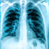
Japan-based mobile communications company Allm has released a collection of COVID-19 pneumonia chest CT images for free through its Join mobile app.
The app enables clinicians to view medical images for various modalities in an embedded DICOM viewer. The updated library includes a set of COVID-19 images created with help from medical institutions in Japan.
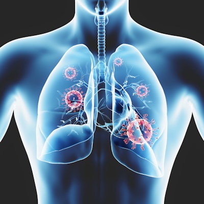
Each case in the COVID-19 library comes with data on patient symptoms and progress, and the company intends to regularly update the image set. Allm created the library as a tool to improve diagnostic accuracy and educate medical staff and students about COVID-19 pneumonia.
Interested healthcare professionals who work at medical institutions in Japan or abroad can request free Join access by emailing [email protected]. Existing users can also request free access by emailing the account with their Join ID/email address, name, medical institution, and department.


![Images show the pectoralis muscles of a healthy male individual who never smoked (age, 66 years; height, 178 cm; body mass index [BMI, calculated as weight in kilograms divided by height in meters squared], 28.4; number of cigarette pack-years, 0; forced expiratory volume in 1 second [FEV1], 97.6% predicted; FEV1: forced vital capacity [FVC] ratio, 0.71; pectoralis muscle area [PMA], 59.4 cm2; pectoralis muscle volume [PMV], 764 cm3) and a male individual with a smoking history and chronic obstructive pulmonary disorder (COPD) (age, 66 years; height, 178 cm; BMI, 27.5; number of cigarette pack-years, 43.2, FEV1, 48% predicted; FEV1:FVC, 0.56; PMA, 35 cm2; PMV, 480.8 cm3) from the Canadian Cohort Obstructive Lung Disease (i.e., CanCOLD) study. The CT image is shown in the axial plane. The PMV is automatically extracted using the developed deep learning model and overlayed onto the lungs for visual clarity.](https://img.auntminnie.com/mindful/smg/workspaces/default/uploads/2026/03/genkin.25LqljVF0y.jpg?auto=format%2Ccompress&crop=focalpoint&fit=crop&h=112&q=70&w=112)