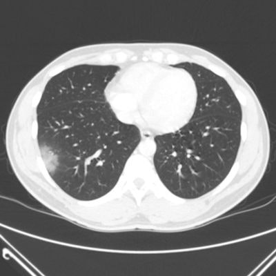
When a new virus eventually named SARS-CoV-2 was identified early in 2020, researchers and clinicians rushed to define its manifestations and determine the best way to diagnose the illness it causes, COVID-19. Chest CT proved to be a valuable tool in those first days, particularly when reverse transcription polymerase chain reaction (RT-PCR) testing wasn't easily available or results were delayed.
Professional organizations such as the American College of Radiology (ACR) made it clear that chest CT should not be used as an initial diagnostic tool for COVID-19. But much research has been published this year suggesting that in specific situations the modality offers a way to quickly assess the severity of disease and triage patients suspected of having the illness.
At the upcoming RSNA 2020 virtual meeting, researchers will share findings from studies that explore the use of CT in the COVID-19 pandemic. To help you prepare, we're offering this overview of key CT research published over the course of this very challenging year for healthcare.
The highlights below indicate just how versatile CT can be when it comes to diagnosing disease and tracking treatment -- and not just for COVID-19: It can be used in applications ranging from lung cancer screening and appendicitis surgery staging to assessing breast cancer patients' cardiovascular disease risk.
COVID-19 concerns
In March, the ACR declared that CT should not be used as a first-line diagnostic or screening tool for COVID-19, and in July, the World Health Organization published guidance regarding the use of chest imaging for the diagnosis and management of COVID-19 patients, emphasizing that CT is suggested for symptomatic patients when RT-PCR testing isn't readily available or when the RT-PCR test is negative but the patient's symptoms point to COVID-19.
But physicians on the front lines of the pandemic continued to argue that chest CT is a useful tool for initial diagnosis of COVID-19 in some clinical situations, citing the low sensitivity of RT-PCR testing and, therefore, the risk of asymptomatic patients infecting others before their illness develops. In January, Chinese researchers reported on the epidemiological and radiological characteristics of this new virus, describing abnormalities on CT that appeared to be indicative of infection; another group found that chest CT could help clarify negative RT-PCR COVID-19 results.
All year, CT helped illuminate more surprising manifestations of COVID-19 and track its transmission. A team from the University of Maryland School of Medicine in Baltimore found that nonchest CT exams performed at the behest of emergency department physicians often also captured pulmonary findings suspicious for COVID-19. In addition, Temple University Health System researchers shared a case of a woman who was diagnosed with the illness when she underwent CT staging for breast cancer.
Chinese investigators from Wuhan, the epicenter of the pandemic, showed that the SARS-CoV-2 virus can transmit from mother to fetus, while a group from Huai'an used the modality to confirm the transmission path of the virus in a cohort of patients who attended the same bathhouse.
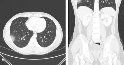 A 33-year-old man presenting with right lower quadrant abdominal pain, found to have acute appendicitis on abdominal/pelvic CT. Axial (left) and coronal (right) views on lung windows demonstrate focal peripheral ground-glass opacity in the right lung base. Images courtesy of the RSNA.
CT has much to offer for long-term tracking of COVID-19 patients, according to Dr. Seth Kligerman, chief of cardiothoracic imaging at the University of California, San Diego.
"CT is helpful in assessing complications from COVID-19, such as pulmonary thromboembolism," he told AuntMinnie.com. "We are also starting to perform follow-up imaging in patients who had severe lung injury due to [COVID-19] and show variable degrees of lung healing. While the lungs in some patients do appear to nearly completely recover, at least on a macroscopic level, others will wind up with permanent fibrosis, which may lead to debilitation."
Imaging economics
Before the peak of the pandemic, CT's use rate had climbed, in contrast to most other modalities, whose rates have tended to stabilize or decrease. Researchers from University of Texas Southwestern Medical Center theorized that this was due in part to hospitals offsetting code bundling with increased CT volumes. A February report from IMV Medical Information Division, a sister company of AuntMinnie.com, confirmed the trend: It showed that CT procedure volume hit an all-time high in 2019, at 91.4 million scans, an uptick that could be due to CT being considered an essential diagnostic tool across a range of applications.
That changed as the pandemic hit and procedure volume dropped -- CT saw a 38% decline, according to an IMV report from late April. In addition, imaging departments faced the challenge of how to keep patients and technologists safe with new sanitation measures that added to exam time and reduced scheduling to avoid patient exposure in waiting rooms.
An imaging workhorse
Beyond COVID-19, CT showed its value this year across a variety of other applications. A research team from Argentina found that using the modality with patients with atypical presentation of appendicitis or negative ultrasound results reduced unnecessary surgery, and a group from Australia found that whole-body CT can be an effective way to image trauma patients.
CT also shed considerable light on how vaping affects patients' lungs and how the practice relates to the development of acute respiratory distress syndrome (ARDS). Another Australian group used CT to document lung disease in coal miners that could have been identified earlier if there had been a robust screening program in place.
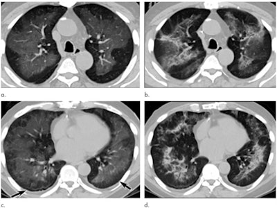 Electronic cigarette or vaping product use-associated lung injury in a 51-year-old man manifesting as an acute lung injury pattern at CT with subsequent organization. (a, c) Axial unenhanced CT images at presentation through (a) mid and (c) lower lungs show ground-glass opacity with subpleural sparing (arrows). (b, d) Axial unenhanced CT images obtained six days later show ground-glass opacity has transitioned to consolidation and mild architectural distortion, consistent with developing organization. The patient was initially treated with antibiotics followed by corticosteroid therapy, with slow clinical improvement. Images and caption courtesy of the RSNA.
On the artificial intelligence side of things, this year a group from Louisiana State University created 3D digitally segmented models using CT data to assess the extent and distribution of COVID-19, and a team of researchers from Wuhan found that a deep-learning algorithm boosted CT's performance for quantifying lung opacification caused by the illness.
More recently, a presentation delivered at the virtual 2020 European Breast Cancer Conference outlined how clinicians used CT scans of coronary artery calcium (CAC) to forecast which patients being treated for breast cancer were at higher risk of developing cardiovascular disease.
| CAC score and risk of cardiovascular disease among women treated for breast cancer | |||||
| Outcome | CAC score | ||||
| 0 | 1-10 | 11-100 | 101-400 | 400+ | |
| Percentage of patients who were hospitalized or died | 5% | 8.9% | 13.5% | 17.5% | 28.3% |
| Hazard ratio (adjusted for age, year) | 1.0 | 1.3 | 1.7 | 2.2 | 3.6 |
Rare results
But CT isn't just for common applications: It also plays a key role in the diagnosis of rare illnesses or conditions. In particular, this past year CT helped clinicians solve the case of a tourist to Cuba who developed symptoms of neurological impairment after her trip. The symptoms were similar to those of U.S. and Canadian diplomats in Cuba in 2016; the diplomats' symptoms were originally thought to be caused by a "sonic weapon" but turned out to be due to pesticides.
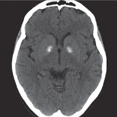 CT scan from a 69-year-old woman with neurological impairment after a trip to Cuba. Findings show bilateral hyperdensity of the globus pallidi. Image courtesy of JAMA Network.
Egyptian investigators used conebeam CT to remove an ectopic third molar and infected cyst from a patient's maxillary sinus, while clinicians from Qatar used CT to identify and remove a 3-cm needle from a patient who swallowed it during a dental procedure. And a team from Phoenix Children's Hospital found that the modality was effective for planning treatment for children with bicycle handlebar injuries.
Leading lung cancer screening
Throughout the year, the conversation about low-dose CT (LDCT) lung cancer screening continued, with researchers weighing in on the questions of who should get it and how often -- and even exploring what types of patients seek it out (spoiler alert: those who have quit smoking and have higher Lung-RADS categories on LDCT).
A group of researchers from Tennessee found that bringing mobile CT lung cancer screening to patients via a customized, climate-controlled bus and portable CT scanner boosted compliance rates. Meanwhile, a team from Florida described two radiomics features on LDCT that show promise for identifying early-stage lung cancer patients who may be at higher risk for poor survival outcomes, which could translate to earlier intervention.
What's next?
As these studies show, CT is a valuable tool in both the clinical and research arenas. So what's coming up for the modality? Our next pre-RSNA CT article includes even more insights on CT's future from a variety of experts.


