Software developer RadLogics has debuted the latest version of its artificial intelligence (AI)-powered COVID-19 CT algorithm.
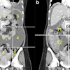
The algorithm stems from a deep-learning process and consists of several models to detect, localize, and segment regions in the lungs infected with COVID-19. In this latest version, three different analyses can now be performed simultaneously on chest CT image scans, including the following:
- A lung's region-of-interest is cropped with lung abnormalities detected.
- The lung lobes are segmented.
- If the nodules plug-in is activated, focal ground-glass opacities (GGOs) are detected.
The measurements are key features in determining if a patient has COVID-19 or not, RadLogics said. The overall system then indicates whether the case is suspected for COVID-19 with a confidence level in percentages.
The algorithm has been validated via an unpublished study led by Dr. Hayit Greenspan from Tel Aviv University and members of the RadLogics team, in which they explored multiple descriptive lung features, including lung and infection statistics, texture, shape, and location. They used the information to train a machine learning-based classifier that distinguishes between COVID-19 and other lung abnormalities.
The study dataset included 2,191 CT cases and demonstrated a 90.8% sensitivity and 85.4% specificity.
The COVID-19 CT software is available worldwide through major OEM distribution partners including Nuance via the AI Marketplace in the U.S. market, RadLogics said.