AI’s use in radiology continues to grow as the technology becomes more available, according to a presentation given September 12 at the International Society for Computed Tomography (ISCT) annual meeting in Boston.
In his talk, Rajiv Gupta, MD, PhD, from Massachusetts General Hospital outlined the state of AI in medical imaging and for CT specialists. He said that AI allows radiologists to view what they cannot see with the naked eye.
“Clearly, there is something to this technology, but there is a lot that still can be done and needs to be done,” Gupta said.
Recently published studies, as well as ongoing research, continue to highlight AI’s potential in clinical use. They suggest that the technology can be used by radiologists as a clinical support tool in making diagnoses and handling time-consuming tasks.
Gupta cited a 2016 talk given by computer scientist Geoffrey Hinton, PhD, where the latter called for radiologists to be trained immediately on how to use the technology. He also predicted that there would be “thousands” of applications for deep learning in health and that deep learning within the next five to 10 years will outperform radiologists stuck in their current imaging techniques.
Fast forward and the number of new radiology job postings has increased from around 1,000 in the first quarter of 2019 to approximately 2,500 job postings in the first quarter of 2022. This is according to an analysis of job board data from the American College of Radiology (ACR).
And regulatory bodies are also getting on board with AI. The number of AI-based medical devices cleared by the U.S. Food and Drug Administration (FDA) has increased sharply from zero in 2010 to 882 as of May 2024. Of these devices, 76% are intended for use by radiologists. A 2023 analysis predicted that an additional 350 new AI products will be cleared by the FDA by the year 2035.
“There are so many products available right now, people are going to marketplaces where you can pick and choose between different types of vendors,” Gupta said.
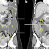
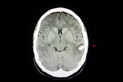
For CT, Gupta said that AI approaches can improve imaging quality and disease prediction. He co-led a study that assessed the performance of a deep learning-based model in predicting intracerebral hemorrhage expansion with dual-energy CT by creating saliency maps. The team found that the convolutional neural network (CNN) model demonstrated promising results. This included an accuracy of 82.7%, 66.7% sensitivity, 75% precision, and 90% specificity.
Gupta also discussed the emerging use of large language models such as ChatGPT and Google Gemini in radiology clinics. These chatbot models are used to aid in doctor-patient communication, record summarization, decision support, and visiting documentation among other uses.
Gupta said he uses the technology routinely on “just about every report I dictate,” adding that his team is piloting a model called Smart Impression.
“This generated impression, using the reports I have dictated, ‘speaks’ in my voice,” Gupta said.
He concluded that whatever deep-learning models interest radiology departments, “all of these” will be supplemented in some way with the goal of making workloads easier to manage.