Newborns' livers can be affected by a variety of congenital and acquired diseases, and imaging plays an important role in the workup and management of these, according to a study published November 7 in RadioGraphics.
But it's crucial to choose the correct imaging modality, noted a team led by Fatemeh Hadian, MBChB, of the Hospital for Sick Children in Toronto, Canada. In a survey of imaging modalities available for this indication, the researchers outlined the pros and cons of using x-ray, ultrasound, MRI, and CT with newborn patients.
"Although they are rare, various congenital and acquired diseases can affect the neonatal liver," the group wrote. "Imaging has [a key] role in the workup and management of many neonatal hepatic abnormalities. For example, imaging is an important component of the workup in neonatal patients with liver failure, helps to narrow the differential diagnosis, and may allow the liver to be salvaged and transplant to be avoided through timely diagnosis of conditions such as neonatal hemochromatosis."
The team outlined the pros and cons of various modalities for neonatal liver imaging:
- X-ray. Although it plays a limited role in evaluating neonatal liver abnormalities, it is often used to assess line placement and bowel abnormalities, the group noted. "Radiographs can demonstrate calcifications in the liver that may be related to neoplastic, vascular, or hepatic parenchymal abnormalities," it wrote.
- Ultrasound. Ultrasound is the primary modality for assessing newborns' livers. Its benefits are that it does not impart radiation, can be performed at bedside, and offers better spatial resolution of the neonatal liver than other modalities. "For assessment of many conditions, [ultrasound] is the only imaging modality required," the authors explained.
- Contrast-enhanced ultrasound. Contrast-enhanced ultrasound is increasingly being used to evaluate liver abnormalities, the team noted. "For focal liver lesions such as hemangiomas, which are the most common lesions in the neonatal liver, assessment with contrast-enhanced US may obviate the need for further investigation with MRI or CT," it wrote.
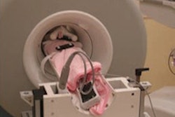
- MRI. When ultrasound assessment is inconclusive, MRI can be used to assess the neonatal liver, and it is required for assessment of neoplasms, some vascular abnormalities, and for diagnosis of hemochromatosis. "MRI can be performed with the 'feed and sleep' technique, which has been reported to be successful for performing diagnostic MRI in greater than 90% of neonates and infants," the authors noted. "In this technique, neonates are fed, bundled, and scanned during natural postprandial sleep."
- CT. CT should be used sparingly because it exposes children to radiation, but it is an option when MR imaging isn't available, according to Hadian and colleagues. "Indications for MRI include suspected acute intra-abdominal bleeding from liver lesions such as hemangiomas, hepatoblastomas, or arteriovenous malformations and liver trauma," they wrote.
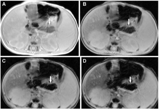
 Neonatal hemochromatosis in a 25-day-old male neonate with neonatal liver failure. Axial MR images of the abdomen with echo times of 2.57 msec (A), 5.36 msec (B), 8.15 msec (C), and 10.94 msec (D) acquired with a 3T MR imaging unit show progressive low signal intensity of the pancreatic parenchyma (arrow) starting at the first echo, suggestive of marked pancreatic siderosis. There is also moderate iron deposition in the liver. Note the sparing of the spleen from siderosis, which is suggestive of neonatal hemochromatosis from gestational alloimmune disease. There is mild splenomegaly. This infant subsequently underwent a liver transplant. Images and caption courtesy of RadioGraphics.
In any case, it's important to keep in mind that since some findings on liver imaging are specific to neonates compared with those of older children, choosing the best modality to image the organ crucial, according to the authors.
"[Selecting] and tailoring the imaging techniques for each indication in neonates is important for optimal care with minimal invasiveness," they concluded.
The complete study can be found here.


