Indications of myosteatosis (that is, the accumulation of fat in skeletal muscles) on preoperative chest CT imaging are linked with worse survival after surgery in patients with stage 0 to IIB non-small cell lung cancer (NSCLC), researchers have reported.
And this result was independent of individuals' skeletal muscle index and subcutaneous adipose tissue, wrote a team led by Lea Mantz, MD, of Massachusetts General Hospital in Boston. The group's findings were published February 11 in Radiology.
"[Our] findings suggest that the myosteatosis markers [skeletal muscle density] and intermuscular adipose tissue] are more important for preoperative risk stratification than the [skeletal muscle index]," the group noted.
Skeletal muscle density and intermuscular adipose tissue are indicators of myosteatosis, the team explained. But the prognostic value of myosteatosis markers in preoperative risk assessment of patients with early-stage NSCLC has not been explored.
Mantz and colleagues investigated whether increased myosteatosis markers found on chest CT imaging before early-stage NSCLC surgery would be linked to and predictive of worse overall survival in these patients. They conducted a study that included 838 patients with stage 0 to IIB NSCLC who underwent lobectomy or bilobectomy at three hospitals between 2014 and 2017. (In Stage 0 NSCLC, the tumor is located only in the top layers of cells lining the air passages, while in Stage IIB NSCLC, the tumor is 5 cm or smaller and disease has spread to the lymph nodes on the same side of the chest.)
Using CT imaging, the authors assessed at patients' 12th thoracic vertebral level the indexes and densities of skeletal muscle, subcutaneous adipose tissue, and intermuscular adipose tissue. Of the total patient cohort, 219 (26%) died after a median follow-up of 5.3 years.
The group reported the following:
- Increased myosteatosis, represented by a decreased skeletal muscle density and increased intermuscular adipose tissue index, was associated with worse overall survival (adjusted hazard ratio, 0.87, or p < 0.001 for skeletal muscle density and 1.24, or p < 0.001 for intermuscular adipose tissue).
- The researchers did not find evidence of an association between overall survival and skeletal muscle index, subcutaneous adipose tissue index, and subcutaneous adipose tissue density.
After myosteatosis marker data from CT imaging was included in preoperative patient assessment, the data's predictive performance of postsurgical outcomes increased from 0.72 (with 1 as reference) to 0.73, the team noted.
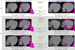
 Body composition analysis of axial CT images at the level of the 12th thoracic vertebral body for two female patients ages (A) 74 and (B) 82 who underwent lobectomy for stage IIB non-small cell lung cancer, both with a body mass index (BMI, calculated as weight in kilograms divided by height in meters squared) of 26.7. The former patient (A) died 2 years after surgery, and the latter patient (B) was censored after 5.2 years. The segmentation results on the right show skeletal muscle (beige), subcutaneous adipose tissue (brown), and intermuscular adipose tissue (IMAT, blue). The anatomic fascia separating the body composition compartments are depicted in red (membranous layer of the superficial fascia) and yellow (fascia transversalis and endothoracic fascia of the fascia trunci interna). Images and caption courtesy of the RSNA.
"We found that worse [overall survival] was associated with lower skeletal muscle density and a higher intermuscular adipose tissue index, independent of other body composition metrics and known confounders," the authors wrote.
The study results suggest that "readily available myosteatosis markers at preoperative chest CT … may improve the preoperative prognostication of patients with early-stage NSCLC," they concluded.
The complete study can be accessed here.