Photon-counting CT (PCCT) is effective and accurate for estimating liver fat fraction compared with MRI proton density fat fraction (PDFF)-based reference standard measurements, researchers have reported.
The findings could offer noninvasive alternatives for assessing liver fat fraction, wrote a team led by Tatjana Dell, MD, of University Hospital Bonn in Germany. The study results were published March 18 in Radiology.
"Given the increasing prevalence of steatotic liver disease and its significant impact on public health in Western societies, histopathologic grading is increasingly reserved for specific cases, such as unclear hepatopathy," the investigators noted. "As a result, a major focus of several medical societies is to advocate for the implementation of noninvasive imaging tools for the detection and monitoring of steatosis."
Fat in the liver, or steatosis, is a critical health problem, the researchers explained. It has typically been assessed by histological assessment via a biopsy, but this strategy is vulnerable to bias due to limitations in biopsy size and not only variabilities in steatosis but also in intra- and interobserver variabilities. MRI PDFF is considered the noninvasive reference standard for diagnosing and quantifying liver steatosis, but PCCT also holds promise for this indication, and as many individuals undergo CT imaging for other reasons, PCCT offers a way to screen opportunistically for fatty liver -- thus improving the prevention and treatment of further liver complications, the authors wrote.
Dell and colleagues evaluated PCCT fat quantification on contrast-enhanced scans, validating the results by comparing them with fat quantification via histopathologic assessment, controlled attenuation parameter (CAP) from transient elastography, and MRI proton density fat fraction (PDFF). Their study included 178 patients with known or suspected liver disease who underwent PCCT between February 2022 and January 2024 (of these, 27 also had liver biopsy, 26 had an ultrasound exam with CAP measurement, and 125 had an MRI exam with a PDFF sequence for hepatic fat fraction quantification). The group measured fat fraction on virtual noncontrast PCCT images using spectral processing software with a three-material decomposition algorithm for fat, liver tissue, and iodine, and graded steatosis for each imaging modality.
The authors reported the following:
- Agreement between PCCT and MRI PDFF assessment of liver fat fraction showed a high intraclass correlation coefficient (ICC) of 0.91.
- In patients with known fibrosis, ICC was 0.84; it was 0.92 in those without the condition.
- Correlation of PCCT-based steatosis grade with histologic and CAP-based steatosis grade was moderate, with Spearman ρ correlation coefficients of 0.65 and 0.45, respectively.
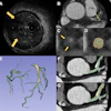
 Liver fat fraction assessment in participants with varying degrees of hepatic steatosis. Fat fraction values were measured in regions of interest (circles) in the right liver lobe on axial contrast-enhanced photon-counting CT fat maps (first row), shown with corresponding proton density fat fraction measurements on axial MRI scans (second row), controlled attenuation parameter measurements from transient elastography (third row), and photomicrographs (hematoxylin-eosin stain; scale bars = 50 μm) of histologic samples (fourth row). Images and caption courtesy of the RSNA.
"[Our study found] excellent agreement of PCCT fat fraction with the noninvasive reference standard, MRI proton density fat fraction (PDFF) … regardless of the presence of underlying fibrosis," the group wrote.
The study results are promising and merit further validation, noted Nikolaos Kartalis, MD, and colleague Aristeidis Grigoiadis, MD, both of the Karolinska University Hospital in Solna, Sweden, in an accompanying commentary.
"Additional prospective studies with larger populations that include additional individuals with healthy livers and individuals with higher degrees of steatosis are needed to identify optimal thresholds and solidify the role of PCCT in the quantification of liver fat content," Kartalis and Grigoiadis wrote.
The complete study can be found here.